Is consciousness to be found in quantum processes in microtubules?

Status
Not open for further replies.
https://science.sciencemag.org/content/369/6504/eaaz2532

The proteasome controls ESCRT-III–mediated cell division in an archaeon

Proteasomal control of division in Archaea
In eukaryotes, proteasome-mediated degradation of cell cycle factors triggers mitotic exit, DNA segregation, and cytokinesis, a process that culminates in abscission dependent on the protein ESCRT-III. By studying cell division in an archaeal relative of eukaryotes, Tarrason Risa et al. identified a role for the proteasome in triggering cytokinesis by an archaeal ESCRT-III homolog. Cell division in this archaeon was driven by stepwise remodeling of a composite ESCRT-III–based division ring, where rapid proteasome-mediated degradation of one ESCRT-III subunit triggered the constriction of the remaining ESCRT-III–based copolymer. These data strengthen the case for the eukaryotic cell division machinery having its origins in Archaea.

Abstract
Proper division of the cell requires coordination between chromosome segregation by the mitotic spindle and cleavage of the cell by the cytokinetic apparatus. Interactions between the mitotic spindle, the contractile ring and the plasma membrane ensure that the cleavage furrow is properly placed between the segregating chromosomes and that new membrane compartments are formed to produce two daughter cells. The microtubule midzone is able to stimulate the cortex of the cell to ensure proper ingression and completion of the cleavage furrow. Specialized microtubule structures are responsible for directing membrane vesicles to the site of cell cleavage, and vesicle fusion is required for the proper completion of cytokinesis.
......

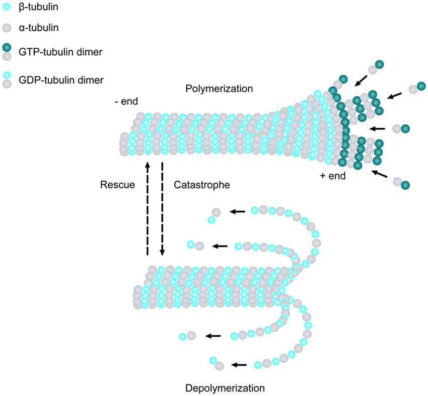
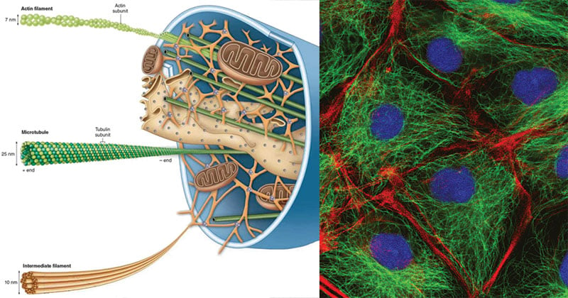
2. Roles of microtubules in cytokinesis
Rearrangement of the microtubule cytoskeleton during mitosis controls the segregation of the chromosomes, the placement of the contractile ring and the completion of cell cleavage.
In animal cells, microtubule-dependent processes in cytokinesis can be divided temporally into two parts. Microtubules of the bipolar spindle dictate the position of the cleavage plane midway between the two asters. Subsequently, microtubules of the spindle midzone promote ingression of the cleavage furrow and the completion of cytokinesis.
To accomplish these tasks, microtubules must interact with the cell cortex to mark the site for cleavage furrow assembly and ultimately direct the assembly of actin and myosin at the furrow region. Microtubules must also communicate with the cell cortex and/or membrane to stimulate the ingression of the cleavage furrow, and they must assemble the midzone and midbody microtubule structures required for the completion of cytokinesis.
....more
https://www.sciencedirect.com/science/article/pii/S0960982200007466#
 
A deep look at a speck of human brain reveals never-before-seen quirks
Extra-strong connections, whorled tendrils and symmetrical cells hint at deep brain mysteries
060421_ls_strong-synapses_feat.jpg

Nerve cells that resided in a woman’s brain send out message-sending tendrils called axons (shown). A preliminary analysis has turned up some super-strong connections between cells.

LICHTMAN LAB/HARVARD UNIVERSITY, CONNECTOMICS TEAM/GOOGLE
By Laura Sanders, JUNE 9, 2021 AT 6:00 AM
A new view of the human brain shows its cellular residents in all their wild and weird glory. The map, drawn from a tiny piece of a woman’s brain, charts the varied shapes of 50,000 cells and 130 million connections between them.
In his intricate map, named H01 for “human sample 1,” represents a milestone in scientists’ quest to provide evermore detailed descriptions of a brain (SN: 2/7/14).
“It’s absolutely beautiful,” says neuroscientist Clay Reid at the Allen Institute for Brain Science in Seattle. “In the best possible way, it’s the beginning of something very exciting.”
Scientists at Harvard University, Google and elsewhere prepared and analyzed the brain tissue sample. Smaller than a sesame seed, the bit of brain was about a millionth of an entire brain’s volume. It came from the cortex — the brain’s outer layer responsible for complex thought — of a 45-year-old woman undergoing surgery for epilepsy. After it was removed, the brain sample was quickly preserved and stained with heavy metals that revealed cellular structures. The sample was then sliced into more than 5,000 wafer-thin pieces and imaged with powerful electron microscopes.
Computational programs stitched the resulting images back together and artificial intelligence programs helped scientists analyze them. A short description of the resulting view was published as a preprint May 30 to bioRxiv.org. The full dataset is freely available online.
060421_ls_strong-synapses_inline1_680.jpg

These two neurons are mirror symmetrical. It’s unclear why these cells take these shapes.
LICHTMAN LAB/HARVARD UNIVERSITY, CONNECTOMICS TEAM/GOOGLE
For now, researchers are just beginning to see what’s there. “We have really just dipped our toe into this dataset,” says study coauthor Jeff Lichtman, a developmental neurobiologist at Harvard University. Lichtman compares the brain map to Google Earth: “There are gems in there to find, but no one can say they’ve looked at the whole thing.”
But already, some “fantastically interesting” sights have appeared, Lichtman says. “When you have large datasets, suddenly these odd things, these weird things, these rare things start to stand out.”
https://www.sciencenews.org/article/brain-map-neurons-connections-google-harvard?
 
A deep look at a speck of human brain reveals never-before-seen quirks
Extra-strong connections, whorled tendrils and symmetrical cells hint at deep brain mysteries
060421_ls_strong-synapses_feat.jpg

Nerve cells that resided in a woman’s brain send out message-sending tendrils called axons (shown). A preliminary analysis has turned up some super-strong connections between cells.

LICHTMAN LAB/HARVARD UNIVERSITY, CONNECTOMICS TEAM/GOOGLE
By Laura Sanders, JUNE 9, 2021 AT 6:00 AM
060421_ls_strong-synapses_inline1_680.jpg

These two neurons are mirror symmetrical. It’s unclear why these cells take these shapes.
LICHTMAN LAB/HARVARD UNIVERSITY, CONNECTOMICS TEAM/GOOGLE
https://www.sciencenews.org/article/brain-map-neurons-connections-google-harvard?

Their environment . Their energy . The nutrients in the cells . Cell wall activity and outside the cell fluid .
 
Their environment . Their energy . The nutrients in the cells . Cell wall activity and outside the cell fluid .
Also known as the cytoskeleton.
What you see are millions of microtubules and filaments. That is what the OP question addresses.
250px-Cellular_Fluid_Content.jpg

The distribution of the total body water in mammals between the intracellular compartment and the extracellular compartment, which is, in turn, subdivided into interstitial fluid and smaller components, such as the blood plasma, the cerebrospinal fluid and lymph

The 67 % is cytoplasm, maintained by microtubules.

Cytoskeleton- Definition, Structure, Functions and Diagram
January 7, 2020 by Sagar Aryal
Cytoskeleton.jpg

Figure: Diagram of Cytoskeleton

A. Microtubules
  • The thickest are the microtubules (20 nm in diameter) which consist primarily of the tubulin protein.
  • Each tubulin subunit is made up of one alpha and one beta-tubulin that are attached to each other, so technically tubulin is a heterodimer, not a monomer. Since it looks like a tube, it is named as microtubule.
  • In a microtubule structure, tubulin monomers are linked both at their ends and along their sides (laterally). This means that microtubules are quite stable along their lengths.
  • Since the tubulin subunits are always linked in the same direction, microtubules have two distinct ends, called the plus (+) and minus (-) ends.
  • On the minus end, alpha-tubulin is exposed, and on the plus end, beta-tubulin is exposed.
  • Microtubules preferentially assemble and disassemble at their plus ends.
Functions
  1. Transportation of water, ions or small molecules.
  2. Cytoplasmic streaming (cyclosis).
  3. Formation of fibers or asters of the mitotic or meiotic spindle during cell division.
  4. Formation of the structural units of the centrioles, basal granules, cilia, and flagella.
https://microbenotes.com/cytoskeleton/
 
continued;
Cellular-cytoskeleton-A-Schematic-of-a-cell-crawling-on-a-2D-substrate-showing-the.png

1: Cellular cytoskeleton. (A) Schematic of a cell crawling on a 2D substrate, showing the prominent locations of the three cytoskeletal components: actin network, microtubules and intermediate filaments. The actin network is denser near the cell membrane aiding in cell migration [10]. B) Fluorescent image of cells with their nuclei (blue) and the cytoskeletal components, actin (red) and microtubules (green). [Wikimedia commons

https://www.researchgate.net/figure...-on-a-2D-substrate-showing-the_fig1_331586446


Note the Centrosome as the central control of the microtubule processes.

Can we say this control organelle is a proto-brain?

Centrosome
In cell biology, the centrosome (Latin centrum 'center' + Greek sōma 'body') (also called cytocentre[1]) is an organelle that serves as the main microtubule organizing center (MTOC) of the animal cell, as well as a regulator of cell-cycle progression. The centrosome provides structure for the cell.
The centrosome is thought to have evolved only in the metazoan lineage of eukaryotic cells.[2]Fungi and plants lack centrosomes and therefore use other structures to organize their microtubules.[3][4]
Although the centrosome has a key role in efficient mitosis in animal cells, it is not essential in certain fly and flatworm species.
400px-Centrosome_%28borderless_version%29-en.svg.png


Functions

Further information: Centrosome cycle

Role of the centrosome in cell cycle progression
Centrosomes are associated with the nuclear membrane during the prophase stage of the cell cycle. During mitosis, the nuclear membrane breaks down, and the centrosome-nucleated microtubules can interact with the chromosomes to build the mitotic spindle.
The mother centriole, the older of the two in the centriole pair, also has a central role in making cilia and flagella.[10]
The centrosome is copied only once per cell cycle, so that each daughter cell inherits one centrosome, containing two structures called centrioles. The centrosome replicates during the S phase of the cell cycle.
During the prophase in the process of cell division called mitosis, the centrosomes migrate to opposite poles of the cell. The mitotic spindle then forms between the two centrosomes. Upon division, each daughter cell receives one centrosome. Aberrant numbers of centrosomes in a cell have been associated with cancer.
Doubling of a centrosome is similar to DNA replication in two respects: the semiconservative nature of the process and the action of CDK2 as a regulator of the process.[16] But the processes are essentially different in that centrosome doubling does not occur by template reading and assembly. The mother centriole just aids in the accumulation of materials required for the assembly of the daughter centriole.
...more
https://en.wikipedia.org/wiki/Centrosome
 
Last edited:
The journey continues

Cytoskeleton and Consciousness: An Evolutionary Based Review Contzen Pereira

ABSTRACT
The fields of quantum biology and physics are now starting to unite to solve the mysteries associated with the field of evolutionary biology. One such question is the origination and propagation of consciousness which has always been ambiguous and in order to understand this concept, many theories have been proposed by several philosophers and scientists.
This review paper agrees with the idea, that evolution is not a random process but hypothesizes, that its succession was managed by the expanding level of consciousness due to cell division and cell differentiation. Several theories propose that the cytoskeleton and its proteins are promoters for consciousness in the brain, which propagates by means of super-conductance.
A better correlation of cytoskeletal evolution and consciousness could help solve the enigma of the origination, propagation and existence of consciousness. This review is a compilation of theories, evidences and scientific studies which intends to bring an association between propagation of consciousness and the evolution of the cytoskeleton and its proteins.
https://www.academia.edu/25505439/Cytoskeleton_and_Consciousness_An_Evolutionary_Based_Review?
 
@ Write4U - You’ve posted about your interest in math as an “explanation” of the physical universe, but do you feel the same about consciousness?
 
@ Write4U - You’ve posted about your interest in math as an “explanation” of the physical universe, but do you feel the same about consciousness?
Interesting, we just touched on that question in Philosophy ; If No Consciousness Exists, By What Right Does The Universe?

I don't think consciousness is a requirement for "intelligent" behavior, and vice versa.

I believe the mathematical essence of spacetime geometry is not conscious, but I call it "quasi intelligent" as is homeostasis in living organisms. The human biome is actually an universe in itself and homeostasis is an unconscious but extremely effective electro-chemical control mechanism that keeps our organs functioning at peak performance, and of course with the help of unconscious bacteria without which we would be unable to convert food into energy.

Only a part of the human biome is conscious, the brain and that relies on sensory date to make a best guess of what's going on out there in the wild yonder.
 
Last edited:
Terrific , Write4U .

And Notice the Physical Essence of your thinking .
TY, most generous of you.

Of course, ultimately everything becomes expressed as observable physical phenomena from simple to complex patterns.

The maths reside in the regularity and consistency of physical expressions.
 
river said:
Terrific , Write4U .

And Notice the Physical Essence of your thinking .


TY, most generous of you.

Of course, ultimately everything becomes expressed as observable physical phenomena from simple to complex patterns.

The maths reside in the regularity and consistency of physical expressions.

Highlighted

Of course . And Maths are not the cause of these regularities nor physical expressions of course as well .
 
Highlighted

Of course . And Maths are not the cause of these regularities nor physical expressions of course as well .
I agree to a point, but it is obvious that when physical events occur in a constant predictable order, there is an underlying logic that is causal for the reliability of these self-ordering processes. I believe these fundamental logical functions are mathematical in nature, Note that I use the term Mathematical as a human invented symbolic language to express logical equations. The universe does not deal with numbers, it deals with "values" and its functional processes are not always random but "algebraic" in essence.
 
river said:
Highlighted

Of course . And Maths are not the cause of these regularities nor physical expressions of course as well .


I agree to a point, but it is obvious that when physical events occur in a constant predictable order, there is an underlying logic that is causal for the reliability of these self-ordering processes. I believe these fundamental logical functions are mathematical in nature, Note that I use the term Mathematical as a human invented symbolic language to express logical equations. The universe does not deal with numbers, it deals with "values" and its functional processes are not always random but "algebraic" in essence.

Hence the periodic table .
 
Proliferation of microtubules in the brain

Light and electron microscopic studies of the distribution of microtubule-associated protein 2 in rat brain: a difference between dendritic and axonal cytoskeletons

R Bernhardt, A Matus

Abstract
A specific antiserum was used to ascertain the distribution of microtubule-associated protein 2 (MAP2) in the rat brain at the light and electron microscope levels. Light microscopy showed MAP2 to be present only in neurons, and only in the dendrites and the perikaryon of each cell.
This same polarized distribution pattern was found in the Purkinje, Golgi, basket, stellate, and granule cells of the cerebellum, and also in neurons of the hippocampus, the olfactory bulb, and the midbrain.
[ quote]While labelling of the dendritic arborization was extensive and intense, MAP2 density tended to decrease in the proximal dendritic trunk. Particularly in large neurons (e.g., Purkinje, Golgi, and pyramidal cells), staining was reproducibly weaker in the cell body than in the main dendrites
[/quote] https://pubmed.ncbi.nlm.nih.gov/6736300/

Lighting Up the Synapses
Author Michael W. Richardson

Dendrites-Synapses.png

Ferreira, et al. The Journal of Neuroscience, 2015.

Dendrites — the arms extending from a neuron’s cell body — receive information from other neurons at sites called synapses. Each dendrite can have thousands of synapses, which together form complex circuits that govern brain function. This image shows a neuron from a mouse hippocampus, an area of the brain responsible for memory, with synapses labelled in yellow and red.
The proper function of these synapses is critical for brain health, and alterations in synapse shape and function are associated with disorders such as autism spectrum disorder, schizophrenia, and Alzheimer's disease. Scientists are studying how synapses are assembled and maintained to provide more information about the underlying cause of these diseases.
https://www.brainfacts.org/brain-an...15/image-of-the-week-lighting-up-the-synapses
 
Appears that microtubules play a role in epigenetics,

An epigenetic regulator emerges as microtubule minus-end binding and stabilizing factor in mitosis

Abstract
The evolutionary conserved NSL complex is a prominent epigenetic regulator controlling expression of thousands of genes. Here we uncover a novel function of the NSL complex members in mitosis. As the cell enters mitosis, KANSL1 and KANSL3 undergo a marked relocalisation from the chromatin to the mitotic spindle. By stabilizing microtubule minus ends in a RanGTP-dependent manner, they are essential for spindle assembly and chromosome segregation.
Moreover, we identify KANSL3 as a microtubule minus-end-binding protein, revealing a new class of mitosis-specific microtubule minus-end regulators. By adopting distinct functions in interphase and mitosis, KANSL proteins provide a link to coordinate the tasks of faithful expression and inheritance of the genome during different phases of the cell cycle.
https://www.ncbi.nlm.nih.gov/pmc/articles/PMC4918316/
 
No. According to the extract you quote, it's the KANSL proteins that do this. They act on microtubules, which play a role in mitosis. No new role for microtubules is suggested.
Thanks for the correction.
I basically wanted to bring attention to the role of microtubules as being instrumental in epigenetics .
It was a new aspect to me.....B-)
 
What is epigenetics
Your genes play an important role in your health, but so do your behaviors and environment, such as what you eat and how physically active you are. Epigenetics is the study of how your behaviors and environment can cause changes that affect the way your genes work. Unlike genetic changes, epigenetic changes are reversible and do not change your DNA sequence, but they can change how your body reads a DNA sequence
Question; How does a "body" read DNA sequence?

The Role of the Microtubule Cytoskeleton in Neurodevelopmental Disorders
Micaela Lasser,
newprofile_default_profileimage_new.jpg
Jessica Tiber and Laura Anne Lowery*
  • Department of Biology, Boston College, Chestnut Hill, MA, United States
Neurons depend on the highly dynamic microtubule (MT) cytoskeleton for many different processes during early embryonic development including cell division and migration, intracellular trafficking and signal transduction, as well as proper axon guidance and synapse formation. The coordination and support from MTs is crucial for newly formed neurons to migrate appropriately in order to establish neural connections. Once connections are made, MTs provide structural integrity and support to maintain neural connectivity throughout development.
Abnormalities in neural migration and connectivity due to genetic mutations of MT-associated proteins can lead to detrimental developmental defects. Growing evidence suggests that these mutations are associated with many different neurodevelopmental disorders, including intellectual disabilities (ID) and autism spectrum disorders (ASD). In this review article, we highlight the crucial role of the MT cytoskeleton in the context of neurodevelopment and summarize genetic mutations of various MT related proteins that may underlie or contribute to neurodevelopmental disorders. Menon and Gupton, 2016; Pacheco and Gallo, 2016; Kirkcaldie and Dwyer, 2017). Newly formed neurons face many challenges as they undergo dramatic changes in shape and migrate their way through the extracellular terrain in order to establish connections with other cells.
Specifically, dynamic MTs play pivotal roles in creating cell polarity, as well as aiding in neural migration in order to establish appropriate neural connectivity throughout development and into adulthood. The elaborate MT network is integral to facilitate numerous morphological and functional processes during neuro-development, including cell proliferation, differentiation and migration, as well as accurate axon guidance and dendrite arborization.
The organization and remodeling of the MT network is also essential for developing neurons to form axons, dendrites and assemble synapses. Moreover, in mature neurons, MTs continue to maintain the structure of axons and dendrites, and serve as tracks for intracellular trafficking, allowing motor proteins to deliver specific cargoes within the cell.

The Role of the Microtubule Cytoskeleton During Neural Development
MTs are one of the major cytoskeletal components present in all eukaryotic cell types. They are composed of α- and β-tubulin heterodimers, which bind to form 13 polarized linear protofilaments that associate laterally together to create the MT (Figure 1; Akhmanova and Steinmetz, 2008). MTs are extremely dynamic structures, existing in either a growing state (polymerization) or shrinking state (depolymerization). The plus ends of MTs can rapidly switch between these two states, going from growth to shrinkage (catastrophe), or from shrinkage to growth (rescue), a process called “dynamic instability” (Mitchison and Kirschner, 1984). Developing neurons depend on this stochastically dynamic nature of the MT cytoskeleton in order to remodel their shape, proliferate and migrate, as well as other processes during different phases of neural development, as described in more detail below.
fncel-12-00165-g001.jpg

Figure 1. Microtubule (MT) basics. MTs are linear structures comprised of α-tubulin and β-tubulin heterodimers. MTs are extremely dynamic, existing in either a growing state (polymerization) or shrinking state (depolymerization), and can rapidly switch from growth to shrinkage (catastrophe) or from shrinkage to growth (rescue). Addition of new GTP-bound heterodimers occurs at the MT plus end during polymerization. Shortly thereafter, the tubulin subunits hydrolyze their bound GTP to GDP. When the addition of GTP-bound heterodimers slows and the MT lattice is composed of predominantly GDP-tubulin, the protofilaments splay apart and the MT depolymerizes.
As brain development relies heavily on proper MT function, defects in the MT cytoskeleton can lead to detrimental effects on neural proliferation, migration and connectivity. Over the last several years, numerous studies have identified mutations within genes coding for proteins that interact with and directly modulate the structure and function of the MT cytoskeleton. Many of these MT-associated mutations have been linked to various neurodevelopmental disorders including lissencephaly, polymicrogyria, autism spectrum disorders (ASD) and intellectual disabilities (ID; Srivastava and Schwartz, 2014; Chakraborti et al., 2016; Stouffer et al., 2016).
The regulation of the MT cytoskeleton during specific stages of brain development still remains an active topic of research. In this review article, we highlight various studies that illustrate important functions of the MT cytoskeleton that contribute to proper neural development and how genetic mutations within MT-related proteins can alter these crucial functions that may lead to disorders of neural development.
.....more
https://www.frontiersin.org/articles/10.3389/fncel.2018.00165/full
 
Neuromorphic Computing
In the field of neuromorphic engineering, researchers study computing techniques that could someday mimic human cognition. Electrical engineers at the Georgia Institute of Technology recently published a "roadmap" that details innovative analog-based techniques that could make it possible to build a practical neuromorphic computer. [9] How does the brain - a lump of 'pinkish gray meat' - produce the richness of conscious experience, or any subjective experience at all?
Scientists and philosophers have historically likened the brain to contemporary information technology, from the ancient Greeks comparing memory to a 'seal ring in wax,' to the 19th century brain as a 'telegraph switching circuit,' to Freud's subconscious desires 'boiling over like a steam engine,' to a hologram, and finally, the computer. [8] Discovery of quantum vibrations in 'microtubules' inside brain neurons supports controversial theory of consciousness. The human body is a constant flux of thousands of chemical/biological interactions and processes connecting molecules, cells, organs, and fluids, throughout the brain, body, and nervous system. Up until recently it was thought that all these interactions operated in a linear sequence, passing on information much like a runner passing the baton to the next runner.
However, the latest findings in quantum biology and biophysics have discovered that there is in fact a tremendous degree of coherence within all living systems. The accelerating electrons explain not only the Maxwell Equations and the Special Relativity, but the Heisenberg Uncertainty Relation, the Wave-Particle Duality and the electron’s spin also, building the Bridge between the Classical and Quantum Theories.
The Planck Distribution Law of the electromagnetic oscillators explains the electron/proton mass rate and the Weak and Strong Interactions by the diffraction patterns. The Weak Interaction changes the diffraction patterns by moving the electric charge from one side to the other side of the diffraction pattern, which violates the CP and Time reversal symmetry. The diffraction patterns and the locality of the self-maintaining electromagnetic potential explains also the Quantum Entanglement, giving it as a natural part of the Relativistic Quantum Theory and making possible to understand the Quantum Biology
https://www.academia.edu/22128506/N...?auto=download&email_work_card=download-paper
 
Status
Not open for further replies.
Back
Top