Is consciousness to be found in quantum processes in microtubules?

Status
Not open for further replies.
continued.....
brain-wired-for-addiction-spotlight.jpg

https://today.tamu.edu/2018/01/05/how-do-synapses-work/

The microtubule cytoskeleton at the synapse
JulieParatoabFrancescaBartolinia

Highlights
Dynamic microtubules enter dendritic spines in an activity-dependent manner, where they contribute to structural plasticity.
The microtubule cytoskeleton acts as a scaffold at inhibitory postsynaptic sites, where it controls the “influx” and “efflux” of receptors during synaptic plasticity.
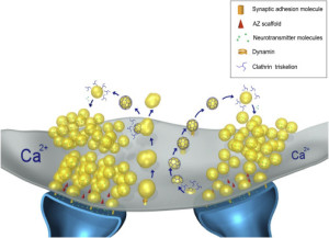
Presynaptic microtubules play multiple roles in bouton organization, local synaptic vesicle trafficking and mitochondrial arrangement in the terminal.
Synaptic microtubule dysfunction may underlie neurological disease.
Abstract
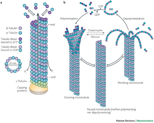
In neurons, microtubules (MTs) provide routes for transport throughout the cell and structural support for dendrites and axons. Both stable and dynamic MTs are necessary for normal neuronal functions. Research in the last two decades has demonstrated that MTs play additional roles in synaptic structure and function in both pre- and postsynaptic elements.
Here, we review current knowledge of the functions that MTs perform in excitatory and inhibitory synapses, as well as in the neuromuscular junction and other specialized synapses, and discuss the implications that this knowledge may have in neurological disease

https://www.sciencedirect.com/science/article/abs/pii/S0304394021002287
 
Last edited:
continued.....

Finally found some information about the actual electrochemical data processing and transport by microtubules. Seems science has not yet been able to sort this out in great detail.

Microtubules
Microtubules are small tubes formed by thirteen filamentous strands. Each filament is composed of a chain of protein called tubulin. Microtubules in a neuron are used to transport substances to different parts of the cell.
For example, neurotransmitters are made in the cell body close to the nucleus, but need to travel long distances to the end of axons where they will be used for
synaptic transmission. Microtubules are the route via which these essential molecules are transported from the site of synthesis to where they are needed.
Advanced
Microtubules, neurofilaments, and microfilaments compose the cytoskeletal elements of a neuron. The earliest accounts of these cellular structures included the writing of not only Ramon y Cajal, the neuroanatomist, but also Freud and Alzheimer in their studies of cellular motility and tubules.
Very recent molecular research has expanded our understanding of these elements of cell architecture, motility, and shape
(Nixon, 1998). Neurons appear to differ in the properties of their cytoskeleton that may reflect functional characteristics, including susceptibility to injury and ability to repair following injury.
Cytoskeletal elements are synthesized in the cell body of a neuron, but delivered throughout the length of the neuron's axon (which composes approximately 99% of the neuron's structure) where they form large molecular assemblies or matrices. Until recently it was believed that the cytoskeletal structure was fairly homogeneous throughout the length of the axon. More recent evidence, however, indicates otherwise.
For example, in mammalian peripheral neurons neurofilament protein content decreases almost 2-fold distally (away from the cell body), while microtubule content increases moving away from the cell body. Neurofilaments collect locally in regions of
myelination in response to signals emanating from the oligodendrocyte. The neurofilament also increases the caliber of the axon underlying myelinated segments of normal neurons.
The dynamic behavior of neuronal filament proteins, including regional accumulation and the formation of cross-links to stabilize matrices, is under the control of protein kinases and phosphatases, enzymes that regulate the molecular activity at the ends of these structural proteins. Our understanding of these enzymes will yield useful information for the treatment of a number of degenerative neurological diseases (Alzheimer's Disease, Lewy body dementia, amyotropic lateral sclerosis) that are characterized by the abnormal accumulation of cytoskeletal elements along the axon
(Julien, 1999; McShea, Wahl & Smith, 1999; Mukaetova-Ladinska, et al., 2000).
https://psych.athabascau.ca/html/Psych402/Biotutorials/1/microtubules.shtml

Figure 11a: Synaptic Transmission


upload_2022-4-5_10-39-15.png

Figure 11a illustrates the site where information is conveyed from one neuron to the next. At this junction, called the synapse, chemicals are used to transmit the electrical neuronal impulse. The structures (magnification approximately 93,000X) and substances involved in synaptic transmission at a directed synapse are identified, and the sequence of events in chemical transmission are described. A directed synapse is one in which the neurotransmitter release sites and receptor sites are close, but not touching. The cleft or space between these structures is typically 0.02-0.05 microns wide. A greater distance between the site of neurotransmitter release and the site of reception characterizes another type of synapse. In this so-called non-directed synapse, the neurotransmitters are released from variscosities (called "string-of-beads") along a neuron's axon.

Note that synapses are the terminal ends of microtubules in axons.

Neuronal Impulse
5. A neuronal impulse is propagated along the axon of the presynaptic neuron to all terminal buttons of that neuron. The rate of neuronal impulses or action potentials reaching the terminal button will determine the amount of neurotransmitter released into the synaptic cleft.
Synapses occur between different neuronal structures, and are named based on the site of neurotransmitter release (presynaptic membrane) and site of receptor binding (postsynaptic membrane). The most common synapses are axodendritic (between axon and dendrite), axosomatic (between axon and soma), or axoaxonic (between two axons).
The basic process of information exchange at axodendritic and axosomatic synapses is the primary topic of this tutorial. Axoaxonic synapses modulate neuronal transmission by either inhibiting or facilitating depolarization at the presynaptic membrane. Dendrodendritic synapses appear to be unique in their ability to transmit in either direction, allowing for reciprocal effects. The multiple modes of communication provided by the varied synaptic connections underlie the complex structure of communication in the nervous system

https://psych.athabascau.ca/html/Psych402/Biotutorials/11/part1.html
 
Last edited:
continued......

How Does A Neuron Stay Polarized
Human Brain, 10.16. 2016

While all cells are complex, the neuron is vastly complex. It is hard to understand how so many different mechanisms all through the cell can be correlated with mental events. Previous posts have clarified very complex processes in both the axons and dendrites.
One post noted how materials are sorted, tagged and transported along the axon to where they are needed.
WC-ART-4-types-of-neurons-208x300.jpg

Different motors and adaptors are required for the movement of varied cargoes.
Another post showed how little is yet known about the extremely rapid complex process of producing, maintaining, secreting and reloading vesicles with neurotransmitters at the synapse. Another post showed that in the dendrite, signaling between the thousands of sections in the arbor is much more complex than previously understood.
Recent research has tried to tackle the way that the neuron is able to keep the environments of the axon and the dendrite so totally different. Research has begun to clarify how axons and dendrites develop, mature and change with neuroplasticity.
Neurons are called polar cells because the functions of the dendrites and axons are so unique. Much of this is based on the different scaffolding and proteins that are enmeshed in the various membranes to provide the electro chemical basis for receiving and sending signals.

Calcium-mechanism-AP-300x217.jpg

The transport of the vesicle along microtubules is another complex system using many different motors—kinesins and dyneins. These move in opposite directions (see post on neuronal transport). These each have multiple adaptor molecules for each type of cargoes. Motor adaptors comprise a large set of different kinds of proteins, not one family. All of this regulated by enzymes that use energy called RABs and ARF.
The microtubules have different electric charges in different directions and these direct some of the traffic to dendrites or axons. While the proteins can be transported back and forth dendrite, material must not go into the axon. Some axonal material goes into the dendrite but not the opposite. Some material is sent into the dendrite where extrusion from the vesicle is rapid and those not put in place are prevented from being released and then lose their polarity.
Much more.......
https://jonlieffmd.com/blog/neuronal-plasticity-blog/how-does-a-neuron-stay-polarized
 
Last edited:
Hmm. What if instead of a human I use a microphone and record some music and don't listen to the recording?

Is there a transfer of "sound information"?
 
Hmm. What if instead of a human I use a microphone and record some music and don't listen to the recording?

Is there a transfer of "sound information"?
Sure in the form of bits and bytes and the recording medium is a HD.
And the "music" itself becomes stored information. If a human listens that information is stored in the pyramidal neurons of the brain and even if later you hear just a segment, you might recall the entire piece including the words.

Recording artists use that instant recall by finding a "hook", a phrase or passage that is "memorable" and becomes an "engram".
Freddy Mercury was a master at communicating a common phrase. That's how he was able to get 10,000 people to sing along with him and Queen.

Interestingly, Queen was an intellectual band.

What did the 4 original members of Queen study?
Education: All four members have attended universities: Mercury had a degree in graphic design and illustration; May has a degree in physics and has done graduate work in astronomy; Taylor has a degree in biology; Deacon earned a degree in electronics with first-class honor
https://www.encyclopedia.com/people/literature-and-arts/music-popular-and-jazz-biographies/queen
 
Last edited:
This is a wonderful experiment in the pentatonic scale to which all people become exposed during their life. There are so many interesting common mental processes in play in this little clip it is truly remarkable.
 
Last edited:
A whole new perspective' Neuro-immune circuits in the brain are responsible for homeostasis.

Microtubules as Regulators of Neural Network Shape and Function: Focus on Excitability, Plasticity and Memory

Abstract

Neuronal microtubules (MTs) are complex cytoskeletal protein arrays that undergo activity-dependent changes in their structure and function as a response to physiological demands throughout the lifespan of neurons. Many factors shape the allostatic dynamics of MTs and tubulin dimers in the cytosolic microenvironment, such as protein–protein interactions and activity-dependent shifts in these interactions that are responsible for their plastic capabilities.
Recently, several findings have reinforced the role of MTs in behavioral and cognitive processes in normal and pathological conditions. In this review, we summarize the bidirectional relationships between MTs dynamics, neuronal processes, and brain and behavioral states. The outcomes of manipulating the dynamicity of MTs by genetic or pharmacological approaches on neuronal morphology, intrinsic and synaptic excitability, the state of the network, and behaviors are heterogeneous. We discuss the critical position of MTs as responders and adaptative elements of basic neuronal function whose impact on brain function is not fully understood, and we highlight the dilemma of artificially modulating MT dynamics for therapeutic purposes
https://www.mdpi.com/2073-4409/11/6/923/htm#
 
Role of microtubules in neuro-transmission.

The microtubule cytoskeleton acts as a key downstream effector of neurotransmitter signaling
Abstract
Microtubules are well known to play a key role in the trafficking of neurotransmitters to the synapse. However, less attention has been paid to their role as downstream effectors of neurotransmitter signaling in the target neuron.
Here, we show that neurotransmitter-based signaling to the microtubule cytoskeleton regulates downstream microtubule function through several mechanisms.
These include tubulin posttranslational modification, binding of microtubule-associated proteins, release of microtubule-interacting second messenger molecules, and regulation of tubulin expression levels.
We review the evidence for neurotransmitter regulation of the microtubule cytoskeleton, focusing on the neurotransmitters serotonin, melatonin, dopamine, glutamate, glycine, and acetylcholine. Some evidence suggests that microtubules may even play a more direct role in propagating action potentials through conductance of electric current. In turn, there is evidence for the regulation of neuro-transmission by the microtubule cytoskeleton.
https://pubmed.ncbi.nlm.nih.gov/20687109/
 
Last edited:
Adressing the quantum portion of the OP.

I wondered why Penrose would be interested in the concept that microtubules might in principle be able to process quantum data. After all microtubules are at nanoscale level and I wondered at what scale quantum processes occur.

At lookie here!

Scale at which quantum effects dominate
When particles are created with dimensions of about 1–100 nanometers, the materials’ properties can change significantly from those at larger scales. This is the size scale where quantum effects can rule the behavior and properties of particles. A fascinating and powerful result of the quantum effects of the nanoscale is the concept of “tunability” of properties. That is, by changing the size of the particle, a scientist can literally fine-tune a material property of interest. At the nanoscale, properties such as melting point, fluorescence, electrical conductivity, magnetic permeability, and chemical reactivity can change as a function of the size of the particle.
https://www.nano.gov/about-nanotechnology/what-is-so-special-about-nano

Microtubule properties seem to meet this description perfectly where applicable.
Microtubules are the largest type of filament, with a diameter of about 25 nanometers (nm), and they are composed of a protein called tubulin.
U3.cp2.2_nrn2631-i1.jpg

figure 1: Microtubules are ever-changing, with reactions constantly adding and subtracting tubulin dimers at both ends of the filament (Figure 1). The rates of change at either end are not balanced — one end grows more rapidly and is called the plus end, whereas the other end is known as the minus end. In cells, the minus ends of microtubules are anchored in structures called microtubule organizing centers (MTOCs). The primary MTOC in a cell is called the centrosome, and it is usually located adjacent to the nucleus.

https://www.nature.com/scitable/topicpage/microtubules-and-filaments-14052932/
 
continued from post# 2349
Do microtubules terminate in the neural synapse where they generate neurotransmitters?

upload_2022-4-6_2-36-25.png

Fig. 1. Schematic of MT functions in two different types of postsynaptic elements. (A) Excitatory postsynaptic site: depolarization of the dendritic spine allows for transient entry of dynamic MTs into the spine. Entry of dynamic MTs into spines has been associated with structural plasticity of the invaded spines. Selective dendritic spine delivery of SytIV is mediated by the MT plus end motor Kif1A. Entry of dynamic MTs into the spine is regulated by the MT plus end binding protein EB3, which can bind to F-actin and F-actin regulators residing in the spine, such as drebrin and cortactin. EB3 is also a binding partner of STIM2, an ER membrane protein and a regulator of Ca2+ dynamics in mushroom spines. This binding may provide an additional pathway for entry of STIM2/smooth endoplasmic reticulum (sER) into the spine. (B) Inhibitory postsynaptic site: the postsynaptic element of inhibitory synapses is typically located directly on the dendrite, cell body or axon hillock. Inhibitory synapses can be glycinergic, GABAergic or mixed. Gephyrin acts as a scaffold protein, anchoring glycine and GABA receptors to the microtubule cytoskeleton. While the lateral diffusion of glycine receptors (GlyRs) in the synapse is affected by F-actin, lateral diffusion outside of the synapse is controlled by MTs, a mechanism that may be important for the dynamic regulation of the neuronal membrane “apparent viscosity” to control the “influx” and “efflux” of receptors at the synapse during synaptic plasticity.
J.

http://www.columbianeuroresearch.or...1/Bartolini-1-s2.0-S0304394021002287-main.pdf
 
Write4U, I see you're trying to understand how the brain stores and processes information. It seems that memory in brains is connected to plasticity . . . perhaps

Rule one is, information is not required to have meaning.
Say the information is a binary string and it's a random string. It might be the result of coin tosses; it might be the output of a Markov process, or an encryption.
How do you tell, if all you have is the string?

Without further meaningful information, you can't tell. The string might be some information that's existed since the BB and that's all you can say about it, without the ancillary input.

But wait, you do have some extra information which conveys meaning. You know about neurons and how they work.
What about how neurons work in parallel, or the cooperative information processing?

This is still mostly research level stuff, no?

What I'm saying is, you need a context in which certain physical things are necessarily fixed, to apply meaning. That might be Rule two, I couldn't really say. . .

This post has been transmitted in order to make a point, about what Information Theory is, and what it isn't.
 
Last edited:
Write4U, I see you're trying to understand how the brain stores and processes information. It seems that memory in brains is connected to plasticity . . . perhaps
Plasticity may be important in "recall". But AFAIK, "storage" it is a process in the pyramidal neurons whereby the data becomes fixed, somewhat like binary data becomes "fixed" on a HD of a computer.

There are 3 individual + 1 communal types of memory and I believe that each memory uses a different type of neuron. Note that when we speak of neurons, we are really talking about the arrays of microtubules that are responsible for data transmission inside the neuron as well as in the cytoplasm and cytoskeleton.
Synaptic plasticity maybe important during the formation and retrieval of information but is not part of the actual information "storage" in the brain's pyramidal neurons which contain stacked microtubules where information is fixed by a process called phosphorylation.

Memory (Encoding, Storage, Retrieval)
1) For most of us, remembering digits relies on
short-term memory, or working memory—the ability to hold information in our minds for a brief time and work with it (e.g., multiplying 24 x 17 without using paper would rely on working memory).
2) Another type of memory is
episodic memory—the ability to remember the episodes of our lives. If you were given the task of recalling everything you did 2 days ago, that would be a test of episodic memory; you would be required to mentally travel through the day in your mind and note the main events.
Semantic memory is our storehouse of more-or-less permanent knowledge, such as the meanings of words in a language (e.g., the meaning of “parasol”) and the huge collection of facts about the world (e.g., there are 196 countries in the world, and 206 bones in your body)
Collective memory refers to the kind of memory that people in a group share (whether family, community, schoolmates, or citizens of a state or a country).
* 3) long term memories also known as "engrams".

Rule one is, information is not required to have meaning.
Say the information is a binary string and it's a random string. It might be the result of coin tosses; it might be the output of a Markov process, or an encryption.
How do you tell, if all you have is the string?
The brain decides what information is "meaningful" by the emotional response it produces. Sometimes an experience is so traumatic that the brain "suppresses" recall.

Other subconscious autonomous neural memory functions are found in the homeostatic neural network in the brain. It controls and regulates all internal chemistry and dynamics that actually keep us alive (with the help of our symbiotic bacterial friends).
Without further meaningful information, you can't tell. The string might be some information that's existed since the BB and that's all you can say about it, without the ancillary input.
Yes, those are the hardwired neural response systems learned at the very beginning of life itself . The "fight or flight" response is already present in single celled neuronless organisms like the Paramecium which has a rudimentary navigational ability. All subsequent responses are just evolved from billions of years of natural selection for survivability.
But wait, you do have some extra information which conveys meaning. You know about neurons and how they work. What about how neurons work in parallel, or the cooperative information processing?
That's why it is called the "neural network", all neurons are in communication with the brain and each other.
This is what is giving rise to terms like "orchestrated", "integrated", "reduction", "composition" in the modern consciousness hypotheses.
This is still mostly research level stuff, no?
Yes, but there is a long history of practical clinical consciousness manipulation by anesthesiology. It is remarkable that we can render a person unconscious but leave the brain's homeostatic neural control system unaffected by the anesthesia. IMO, this may offer some interesting insights later on in the fledgling neural research of the brain.
What I'm saying is, you need a context in which certain physical things are necessarily fixed, to apply meaning. That might be Rule two, I couldn't really say. . .
I agree, but to decipher some 3 billion years of evolution from Prokaryotic organisms to Eukaryotic organisms and the development of electro-chemical neural systems from primarily chemical response systems is undoubtedly going to take a while....!
This post has been transmitted in order to make a point, about what Information Theory is, and what it isn't.
There are several current research approaches :

Orchestrated Objective Reduction (ORCH OR)
https://en.wikipedia.org/wiki/Orchestrated_objective_reduction

Higher Order Syntactic Thought Theory (HOST)
https://www.frontiersin.org/articles/10.3389/fpsyg.2020.00655/full

Memory Consolidation)
https://www.karger.com/Article/Fulltext/363702

Integrated information theory (IIT, Phi)
https://en.wikipedia.org/wiki/Integrated_information_theory

Revisiting the Global Workspace ( orchestrating the hierarchical organisation of the human brain)
https://medicalxpress.com/news/2021-01-revisiting-global-workspace-orchestrating-hierarchical.html
 
Last edited:
More specifically. Hameroff explains how the actual storage process unfolds.

Scientists claim brain memory code cracked
Date: March 9, 2012
Source: Center for Consciousness Studies University of Arizona, Anesthesiology
The standard experimental model for neuronal memory is long term potentiation (LTP) in which brief pre-synaptic excitation results in prolonged post-synaptic sensitivity. An essential player in LTP is the hexagonal enzyme calcium/calmodulin-dependent protein kinase II (CaMKII).
Upon pre-synaptic excitation, calcium ions entering post-synaptic neurons cause the snowflake-shaped CaMKII to transform, extending sets of 6 leg-like kinase domains above and below a central domain, the activated CaMKII resembling a double-sided insect. Each kinase domain can phosphorylate a substrate, and thus encode one bit of synaptic information. Ordered arrays of bits are termed bytes, and 6 kinase domains on one side of each CaMKII can thus phosphorylate and encode calcium-mediated synaptic inputs as 6-bit bytes. But where is the intra-neuronal substrate for memory encoding by CaMKII phosphorylation? Enter microtubules.
Using molecular modeling, Craddock et al reveal a perfect match among spatial dimensions, geometry and electrostatic binding of the insect-like CaMKII, and hexagonal lattices of tubulin proteins in microtubules. They show how CaMKII kinase domains can collectively bind and phosphorylate 6-bit bytes, resulting in hexagonally-based patterns of phosphorylated tubulins in microtubules. Craddock et al calculate enormous information capacity at low energy cost, demonstrate microtubule-associated protein logic gates, and show how patterns of phosphorylated tubulins in microtubules can control neuronal functions by triggering axonal firings, regulating synapses, and traversing scale.
Microtubules and CaMKII are ubiquitous in eukaryotic biology, extremely rich in brain neurons, and capable of connecting membrane and cytoskeletal levels of information processing. Decoding and stimulating microtubules could enable therapeutic intervention in a host of pathological processes, for example Alzheimer's disease in which microtubule disruption plays a key role, and brain injury in which microtubule activities can repair neurons and synapses
https://www.sciencedaily.com/releases/2012/03/120309103701.htm
 
So is there life in the quantum realm ?
That definition is too general.
Life is a pattern formed within the quantum realm. Life emerged from the quantum realm.
Just as consciousness is a pattern formed within the physical realm. Consciousness emerged from the physical realm.
 
There are several current research approaches :
I'm sure there are. Brain information is not all that well understood yet.

I recall reading something on the subject of cooperative activity between neurons, it might have been in SciAm.

Anyhoo, the research found that groups of neurons set themselves up by more or less singing to each other. Those that sing the same synchronous tune then engage in some higher level activity.

These groups or "choirs" of neurons and presumably other support cells aren't fixed by other than the interneuron signalling. That is, the groups emerge (and then cooperate) from what appears to be random, chaotic neural activity.

The random background though is probably some threshold of say, conscious thought.
 
I'm sure there are. Brain information is not all that well understood yet.
I agree and have commented on the lack of coordinated research between the different areas of neurology.
I recall reading something on the subject of cooperative activity between neurons, it might have been in SciAm.
Yes, and I suspect this is a result of "quorum sensing", which is what bacteria use to communicate.
Anyhoo, the research found that groups of neurons set themselves up by more or less singing to each other. Those that sing the same synchronous tune then engage in some higher level activity.
Yes, this seems to be fundamental, considering that in addition to neural communication, all the cells in the body seem to communicate with each other .
These groups or "choirs" of neurons and presumably other support cells aren't fixed by other than the interneuron signalling. That is, the groups emerge (and then cooperate) from what appears to be random, chaotic neural activity.
Apparently when the communication is complimentary it seems to create new synapses and reinforce the communication network. The body has no skills in circuit design so all of that appears random, but it is obviously effective.
The random background though is probably some threshold of say, conscious thought.
To me that seems an entirely reasonable assumption.

I had the experience of standing under a high power transmission tower with a neon light bulb and have it light up. It was awesome.
main-qimg-d3aceea8bc088b6bdb6c6da1aac557f8-pjlq

Now consider a similar micro effect among trillions of microtubules and synapses in the brain!
 
A little additional info on MT

Microtubules as One-Dimensional Crystals: Is Crystal-Like Structure the Key to the Information Processing of Living Systems?
Abstract:
Each tubulin protein molecule on the cylindrical surface of a microtubule, a fundamental element of the cytoskeleton, acts as a unit cell of a crystal sensor. Electromagnetic sensing enables the 2D surface of microtubule to act as a crystal or a collective electromagnetic signal processing system.
We propose a model in which each tubulin dimer acts as the period of a one-dimensional crystal with effective electrical impedance related to its molecular structure. Based on the mathematical crystal theory with one-dimensional translational symmetry, we simulated the electrical transport properties of the signal across the microtubule length and compared it to our single microtubule experimental results. The agreement between theory and experiment suggests that one of the most essential components of any Eukaryotic cell acts as a one-dimensional crystal.
1. Introduction:
In the past, several theoretical studies were completed by physicists on the vibrational characteristics of bio-living systems, which are currently becoming more attractive research domains in the present. State-of-the-art tools are used to detect vibrational aspects and principle authentication. Traditional theoretical methods are not sufficient to understand the process of biological systems [1,2].
The cooperative interaction between individual components of the developed dynamic system of the biological system changes in a specific fashion [3]. Biological materials show complex vibrational patterns. In other words, solid structures have a crystalline structure and the translational symmetry/spatial periodicity confirms them as an integrated mechanical system, which is well known in solid-state physics or lattice theory. The lattice, known as the base, can be found by the duplication of building elements [4]. The electrons interact with the atomic lattice with atomic potentials.
The electronic bandgap is generated when an electronic state does not exist for a certain energy range [5].
Consequently, a wide range of man-made systems is inspired by the design and organization of such biological systems, which include some of their properties. Whereby we have photonic crystals (crystals in which phonons interact with the periodic variation of elasticity and mass of structure elements) and phononic crystals (crystals in which photons are affected by the variation of the dielectric constant of structure) [6,7].
5.3. Electrical Transport Measurements
MT transmittance was measured with a four-probe circuit [21], there are some special frequencies where the microtubule conducts the AC signal with almost no AC resistance (<<<0.04 MΩ) at those frequencies.
The details can be found in [21]. Both spectra were measured between 10 kHz to 20 GHz.
It has been suggested that the microtubule resonance frequencies significantly change the lattice geometry of the microtubule. Every single resonance frequency is associated with particular lattice geometry. Using a specially designed scanning tunneling microscope, the cylindrical surface has been imaged live.
Several fundamentally different experiments support each other in the finding that the microtubule is an insulator; however, several interesting properties of microtubule emerge as soon as it is triggered by resonance frequencies. Using several versions of the tubulin proteins, it has been demonstrated that electromagnetic resonance has a profound effect, even in their self-assembly.
......more

https://www.mdpi.com/2073-4352/11/3/318/htm
 
Status
Not open for further replies.
Back
Top