Is consciousness to be found in quantum processes in microtubules?

Status
Not open for further replies.
Yeah, unfortunately mdpi isn't a trustworthy source of scientific publications.
It's more a predatory site.
And on what evidence do you make this vague slander?
Is anything in that article bad science and false?

If it is an unreliable site, I'll drop it from my list . But this is the first time I hear something negative from this site.
Show me a peer review?
 
Last edited:
As I understand it, consciousness is an excitation of a "field" generated by the brain .

EMF transmitters and receivers in the brain
It has been known since the 19th century that the brain generates its own EM field, which can be detected by electrodes inserted to the brain. Its source is electrical dipoles within the neuronal membranes caused by the motion of ions in and out of those membranes during action potentials and synaptic potentials.
The periodic discharge of neurons—firing or action potentials—generates EMF waves that propagate out of the neuron and into the surrounding inter-neuronal spaces where they overlap and combine to generate the brain’s global EM field that is routinely measured by brain scanning techniques such as electroencephalography (EEG) and magnetoencephalography (MEG). The human brain, therefore, possesses around 100 billion EMF transmitters.
The human brain also possesses at least 100 billion EMF receivers as each neuron is bounded by a membrane embedded with thousands of voltage-gated ion channels whose firing is triggered by EM field fluctuation across the membrane.
Note that the author generalizes the function of the "neural network" without mentioning the role of microtubules that comprise the billions of EMF transmitters.
Although these channels are generally assumed to respond only to large fluctuations of tens of millivolts across the membrane, much larger than the global EM field strength, EM field potential changes of less than 1 mV across the neuronal membrane are nevertheless capable of modulating neuronal firing (Schmitt et al. 1976).
Moreover, for neurons poised close to the critical firing potential, the opening of just a single ion channel may be sufficient to trigger firing (Arhem and Johansson 1996). This degree of sensitivity suggests that very tiny changes in membrane potential, of similar strength to spontaneous fluctuations in the brain’s endogenous EM field, may influence the firing of neurons that are already close to firing.
I recall an objection voiced by a poster that if an article does not identify microtubules by name, it may be assumed that microtubules do not play a role in the process. Clearly, this is not a valid argument, because it depends on the level of research and "sufficient narrative information".
The fact is that when we speak of the neural network we are by physical necessity including "microtubules" as the actual transport mechanism, much as "copper wire" is the transport mechanism in the electrical network of a residence.

The cemi field theory of consciousness
The conscious electromagnetic information (cemi) field theory claims that the brain’s EM field is the physical substrate of consciousness. It was first outlined in a book published in 2000 in which I proposed that the brain’s ‘EM field … integrates information from all of the calculations … performed by … [its] logic gates (McFadden 2000).
.....more
The idea that the seat of consciousness is simply the brain’s EM field may initially sound outlandish but is no more extraordinary than the claim that the seat of consciousness is the matter of the brain.
All it involves is going from the right to the left hand side of Einstein’s famous equation, E = mc2 thereby replacing the notion that consciousness is encoded by matter of the brain, with that of proposing that it is encoded by the energy of the EM fields generated by the motions of its charged matter. (Note that, by illustrating this idea with Einstein’s equation, I am not, of course, proposing any interconversion of matter and energy in the brain.)
Matter and energy are equally physical; but, instead of being composed of material, the cemi field theory proposes that our thoughts are composed of the brains EM field energy. This is a kind of dualism, but it is scientific dualism based on the physical difference between matter and energy, rather than a metaphysical distinction between matter and spirit.
.........more
https://academic.oup.com/nc/article/2020/1/niaa016/5909853#227499907

I hope this is a more reliable source?

If you do a search for microtubules, you get zero (0) results. I can not imagine how this research facility connected with Oxford University has managed to escape noticing the role microtubules play in the "neural network"

https://academic.oup.com/nc/search-..._SiteID=5412&SearchSourceType=1&allJournals=1

Is this a result of fracture due to scientific specialization?
 
Last edited:
If a "thought" is an excitation in the EM field of the brain, would it be experienced as an internal hologram?

Holography
Holography is a technique that enables a wavefront to be recorded and later re-constructed. Holography is best known as a method of generating three-dimensional images, but it also has a wide range of other applications. In principle, it is possible to make a hologram for any type of wave.
220px-Holomouse2.jpg
Two photographs of a single hologram taken from different viewpoints
A hologram is made by superimposing a second wavefront (normally called the reference beam) on the wavefront of interest, thereby generating an interference pattern which is recorded on a physical medium. When only the second wavefront illuminates the interference pattern, it is diffracted to recreate the original wavefront. Holograms can also be computer-generated by modelling the two wavefronts and adding them together digitally. The resulting digital image is then printed onto a suitable mask or film and illuminated by a suitable source to reconstruct the wavefront of interest.
https://en.wikipedia.org/wiki/Holography

This seems to suggest that human ability for visual triangulation would allow the brain to create an internal hologram which can be experienced as an "observation"

Does (can) human visual abilities create a holographic image in the brain? Can we simulate it with known holographic photography?

Holographic augmented reality based on three-dimensional volumetric imaging for a photorealistic scene

3. Hologram rendering
Acquiring a 3D volumetric point cloud of an object to generate holograms has several advantages. 1) For a holographic AR service, object information is needed from various viewpoints simultaneously. 3D volumetric data containing all viewpoint information is a good alternative to satisfy this condition. 2) A high-quality hologram can be generated by densifying object points located in a 3D space using various spatial techniques.
3) As the normal of each object point can be calculated in the process of obtaining the object point, the angle of the reflected wave reaching the hologram plane can be calculated. If the angle of the reflected light is known, the intensity of interference on the hologram plane can be realistically calculated. Therefore, a more realistic hologram can be generated, and high-quality reconstruction results can be obtained. We divide the process into 3D volumetric model integration and hologram generation for AR.

getImage.cfm


https://opg.optica.org/oe/fulltext.cfm?uri=oe-28-24-35972&id=442589
 
What do neurons process? What do synapses process? What process takes place during mitosis?
What information does the cytoplasm and cytoskeleton of every living cell in a body process?
Rhetorical questions are all well and good, but I asked you a direct question. Here it is again:

What exactly do they process? A process involves operating on an input to produce an output. What is the input, output and processing of a microtubule?
I think that in your rush to spam more random factoids and speculations about microtubules, you lost track of what I asked you.

If there's an answer buried somewhere in your latest wall of text and pretty pictures, can you please dig it out for me and create a one paragraph summary that attempts to answer the questions I asked you?

Thanks.
 
W4U said:
What do neurons process? What do synapses process? What process takes place during mitosis?
What information does the cytoplasm and cytoskeleton of every living cell in a body process?
Rhetorical questions are all well and good, but I asked you a direct question. Here it is again:

What exactly do they process? A process involves operating on an input to produce an output. What is the input, output and processing of a microtubule?
I think that in your rush to spam more random factoids and speculations about microtubules, you lost track of what I asked you.

If there's an answer buried somewhere in your latest wall of text and pretty pictures, can you please dig it out for me and create a one paragraph summary that attempts to answer the questions I asked you?

Thanks.
Perhaps in your haste to offer constructive critique you are overlooking the comprehensive answer I gave you.
"Do you know what neurons process?"

If you do then I don't need to tell you what microtubules process. Microtubules are the data transport mechanism of neurons, just as a copper wire is the electrical transport system of power cables.

As to cytoplasm and cytoskeleton, every description of data transport between cells involves microtubules. I do not need to provide a list of specifics.

ALL electrochemical data-transport throughout the body is processed by microtubules.
That's what they do! They are the highways along which all data is transported and distributed, from your toes to your brain. Dynein and Kinesin are the trucks that transport the data.

Microtubules do not need neurons at all. Neurons need microtubules. Neurons are specialised cells, but every single cell of the body communicates with other cells via their microtubules. Microtubules allow cells to communicate whatever they communicate.

ALL of it! You may know these processes by different names , but all these specialized names involve microtubules.

ALL CELLS CONTAIN MICROTUBULES AS THEIR INTRACELLULAR AND INTERCELLULAR DATA TRANSPORT PROCESSORS.

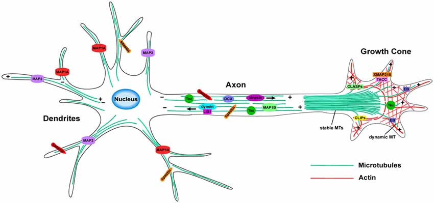
Microtubule Motors and Movements
Microtubules are responsible for a variety of cell movements, including the intracellular transport and positioning of membrane vesicles and organelles, the separation of chromosomes at mitosis, and the beating of cilia and flagella. As discussed for actin filaments earlier in this chapter, movement along microtubules is based on the action of motor proteins that utilize energy derived from ATP hydrolysis to produce force and movement. Members of two large families of motor proteins—the kinesins and the dyneins—are responsible for powering the variety of movements in which microtubules participate.
Go to:
Identification of Microtubule Motor Proteins
Kinesin and dynein, the prototypes of microtubule motor proteins, move along microtubules in opposite directions—kinesin toward the plus end and dynein toward the minus end (Figure 11.45).
The first of these microtubule motor proteins to be identified was dynein, which was isolated by Ian Gibbons in 1965. The purification of this form of dynein (called axonemal dynein) was facilitated because it is a highly abundant protein in cilia, just as the abundance of myosin facilitated its isolation from muscle cells.
The identification of other microtubule-based motors, however, was more problematic because the proteins responsible for processes such as chromosome movement and organelle transport are present at comparatively low concentrations in the cytoplasm. Isolation of these proteins therefore depended on the development of new experimental methods to detect the activity of molecular motors in cell-free systems.
ch11f45.jpg

Figure 11.45
Microtubule motor proteins. Kinesin and dynein move in opposite directions along microtubules, toward the plus and minus ends, respectively. Kinesin consists of two heavy chains, wound around each other in a coiled-coil structure, and two light chains. (more...)
Organelle Transport and Intracellular Organization
One of the major roles of microtubules is to transport membrane vesicles and organelles through the cytoplasm of eukaryotic cells. As already discussed, such cytoplasmic organelle transport is particularly evident in nerve cell axons, which may extend more than a meter in length. Ribosomes are present only in the cell body and dendrites, so proteins, membrane vesicles, and organelles (e.g., mitochondria) must be transported from the cell body to the axon.
Via video-enhanced microscopy, the transport of membrane vesicles and organelles in both directions can be visualized along axon microtubules, where kinesin and dynein carry their cargoes to and from the tips of the axons, respectively. For example, secretory vesicles containing neurotransmitters are carried from the Golgi apparatus to the terminal branches of the axon by kinesin. In the reverse direction, cytoplasmic dynein transports endocytic vesicles from the axon back to the cell body.
Microtubules similarly transport membrane vesicles and organelles in other types of cells. Because microtubules are usually oriented with their minus end anchored in the centrosome and their plus end extending toward the cell periphery, different members of the kinesin and dynein families are thought to transport vesicles and organelles in opposite directions through the cytoplasm (Figure 11.46).
Conventional kinesin and other plus end-directed members of the kinesin family carry their cargo toward the cell periphery, whereas cytoplasmic dyneins and minus end-directed members of the kinesin family transport materials toward the center of the cell.
In addition to transporting membrane vesicles in the endocytic and secretory pathways, microtubules and associated motor proteins position membrane-enclosed organelles (such as the endoplasmic reticulum, Golgi apparatus, lysosomes, and mitochondria) within the cell. For example, the endoplasmic reticulum extends to the periphery of the cell in association with microtubules
https://www.ncbi.nlm.nih.gov/books/NBK9833/
 
Last edited:
And on what evidence do you make this vague slander?
A quick Google will turn up quite bit of controversy about it being a predatory source.

Whether or not it is, arfa brane has every right to not trust it without independent corroboration.
 
continued.....

A special type of data transport may be found in mitosis;

Separation of Mitotic Chromosomes
As discussed earlier in this chapter, microtubules reorganize at the beginning of mitosis to form the mitotic spindle, which plays a central role in cell division by distributing the duplicated chromosomes to daughter nuclei. This critical distribution of the genetic material takes place during anaphase of mitosis, when sister chromatids separate and move to opposite poles of the spindle.
Chromosome movement proceeds by two distinct mechanisms, referred to as anaphase A and anaphase B, which involve different types of spindle microtubules and associated motor proteins.
Anaphase A consists of the movement of chromosomes toward the spindle poles along the kinetochore microtubules, which shorten as chromosome movement proceeds (Figure 11.48). This type of chromosome movement appears to be driven principally by kinetochore-associated motor proteins that translocate chromosomes along the spindle microtubules in the minus end direction, toward the centrosomes.
Cytoplasmic dynein is associated with kinetochores and may play a role in poleward chromosome movement, as may minus end-directed members of the kinesin family. The action of these kinetochore motor proteins is coupled to disassembly and shortening of the kinetochore microtubules, which may be mediated by some members of the kinesin family that act as microtubule-destabilizing enzymes.

ch11f48.jpg

Figure 11.48
Anaphase A chromosome movement. Chromosomes move toward the spindle poles along the kinetochore microtubules. Chromosome movement is thought to be driven by minus end-directed motor proteins associated with the kinetochore. The action of these motor proteins (more...)
Anaphase B refers to the separation of the spindle poles themselves (Figure 11.49). Spindle-pole separation is accompanied by elongation of the polar microtubules and is similar to the initial separation of duplicated centrosomes to form the spindle poles at the beginning of mitosis (see Figure 11.43). During anaphase B the overlapping polar microtubules slide against one another, pushing the spindle poles apart.
This type of movement has been found to result from the action of several plus end-directed members of the kinesin family, which crosslink polar microtubules and move them toward the plus end of their overlapping microtubule—away from the opposite spindle pole. In addition, the spindle poles may be pulled apart by the astral microtubules.
The mechanism responsible for this type of movement has not been established, but it could result from the action of cytoplasmic dynein anchored to the cell cortex or another structure in the cytoplasm. The translocation of such an anchored dynein motor along astral microtubules in the minus end direction would have the effect of pulling the spindle poles apart, toward the periphery of the cell. Alternatively, a motor protein associated with the spindle poles could move along astral microtubules in the plus end direction, which would also pull the spindle poles toward the cell periphery.
https://www.ncbi.nlm.nih.gov/books/NBK9833/
 
Perhaps in your haste to offer constructive critique you are overlooking the comprehensive answer I gave you.
As far as I can tell, you're yet to give an answer.
If you do then I don't need to tell you what microtubules process. Microtubules are the data transport mechanism of neurons, just as a copper wire is the electrical transport system of power cables.
Data transport is not the same thing as data processing.

Your claim is that microtubules are processors. Please try to support that.

What is their input? What processing do they do? What outputs do they produce, and how do they differ from the input?
 
As far as I can tell, you're yet to give an answer.

Data transport is not the same thing as data processing.

Your claim is that microtubules are processors. Please try to support that.

What is their input? What processing do they do? What outputs do they produce, and how do they differ from the input?
Well we are making progress. At least we come to the admission that microtubules transport data.
As to what and how microtubules process data, don't ask me, ask these people:

Information processing in brain microtubules
Abstract
Models of the mind are based on the idea that neuron microtubules can perform computation. From this point of view, information processing is the fundamental issue for understanding the brain mechanisms that produce consciousness. The cytoskeleton polymers could store and process information through their dynamic coupling mediated by mechanical energy. We analyze the problem of information transfer and storage in brain microtubules, considering them as a communication channel. We discuss the implications of assuming that consciousness is generated by the subneuronal process.
https://www.sciencedirect.com/science/article/abs/pii/S0303264705000912
 
Continuing with defining microtubule processes.

Bundles of Brain Microtubules Generate Electrical Oscillations

Abstract
Microtubules (MTs) are long cylindrical structures of the cytoskeleton that control cell division, intracellular transport, and the shape of cells. MTs also form bundles, which are particularly prominent in neurons, where they help define axons and dendrites.
MTs are bio-electrochemical transistors that form nonlinear electrical transmission lines. However, the electrical properties of most MT structures remain largely unknown. Here we show that bundles of brain MTs spontaneously generate electrical oscillations and bursts of electrical activity similar to action potentials. Under intracellular-like conditions, voltage-clamped MT bundles displayed electrical oscillations with a prominent fundamental frequency at 39 Hz that progressed through various periodic regimes. The electrical oscillations represented, in average, a 258% change in the ionic conductance of the MT structures.
Interestingly, voltage-clamped membrane-permeabilized neurites of cultured mouse hippocampal neurons were also capable of both, generating electrical oscillations, and conducting the electrical signals along the length of the structure.
Our findings indicate that electrical oscillations are an intrinsic property of brain MT bundles, which may have important implications in the control of various neuronal functions, including the gating and regulation of cytoskeleton-regulated excitable ion channels and electrical activity that may aid and extend to higher brain functions such as memory and consciousness.
....
MTs are thought to generate oscillatory electric fields at expense of elasto-electrical vibrations12, which may explain our findings that electrically-stimulated MTs behave as biological transistors behaving as sophisticated nonlinear transmission lines, capable of supporting the amplification and axial transfer of electrical signals13,14,15,16,17,18. Within the cytoplasm MT-generated variable currents may contribute to the presence and modulation of large intracellular electric fields, which in turn, will help control cell function.
The above arguments support a potentially relevant role of electrical oscillations on brain MT bundles, which should be critical to neural function. We recently reported that MT sheets sustain electrical oscillations19, which are driven by a permanent electrical polarization from local asymmetries in the ionic distributions between the intra- and extra-MT environments. Thus, the MT wall behaves as an electrical oscillator that produces oscillatory ionic currents with variable amplitude and periodicity depending on the driving force and ionic compositions, and is consistent with the periodic on-off switching of the nanopores.
In this context, here we explored whether rat brain MT bundles do have electrical properties consistent with electrical oscillators. Our findings indicate that rat brain MT bundles generate a wide variety of endogenous electrical oscillations. Interestingly, the cytoskeleton of cultured adult mouse hippocampal neurons also supported electrical oscillations establishing an electrical role for brain MTs.
much more detailed description of microtubule processes ................
https://www.nature.com/articles/s41598-018-30453-2
 
Well we are making progress. At least we come to the admission that microtubules transport data.
I'm not sure that they do. What data do they transport? What does the transporting, exactly? How is the data encoded?
As to what and how microtubules process data, don't ask me, ask these people:
I thought it was your claim that microtubules are data processors. Are you now saying you're not sure about that? Or are you saying you think they are but you don't know why? If it's the latter, what makes you think they are, if you don't have a clue what the mechanism is?
Ok. Let's see...

"Models of the mind are based on the idea that neuron microtubules can perform computation. From this point of view, information processing is the fundamental issue for understanding the brain mechanisms that produce consciousness."​

It sounds like they are assuming that microtubules can perform computation. Then, given the assumption that this is true, they are thinking about the implications for "models of the mind".

I'm not asking about assumptions, though. I'm asking whether there's any evidence that microtubules perform computations.

"The cytoskeleton polymers could store and process information through their dynamic coupling mediated by mechanical energy."​

Could? Well, do they or don't they? You're not sure?

"We analyze the problem of information transfer and storage in brain microtubules, considering them as a communication channel."​

Okay. So, I'm interested in the conclusions they reach about his. Is the problem solved, or not? Are microtubules a "communication channel" or aren't they? And what about the processing I asked about? Is there any?

"We discuss the implications of assuming that consciousness is generated by the subneuronal process."​

They discuss the implications of an assumption. Okay.

It sounds like their claims are much more circumscribed than your claims, Write4U. Do you agree?
 
I thought it was your claim that microtubules are data processors. Are you now saying you're not sure about that? Or are you saying you think they are but you don't know why? If it's the latter, what makes you think they are, if you don't have a clue what the mechanism is?
I think they are because the scientists tell me they are. That is why I quote them lest there be any misunderstanding.
Okay. So, I'm interested in the conclusions they reach about his. Is the problem solved, or not? Are microtubules a "communication channel" or aren't they? And what about the processing I asked about? Is there any?
Who are you talking to? You are reading what they have to say, no? I am merely quoting the language used by bona fide scientists. If you have objections talk to them. I only report on the current state of the science.
"We discuss the implications of assuming that consciousness is generated by the subneuronal process."
They discuss the implications of an assumption. Okay.
Seems to me they are discussing the various models based on the assumption that the microtubules is the only candidate that meets the minimum requirements of the models.
It sounds like their claims are much more circumscribed than your claims, Write4U. Do you agree?
No I don't . I believe that the various discussions confirm the initial assumptions. I believe that the quoted passages speak volumes about the electrical and chemical information that microtubules process and transport. I purposely do not clutter the pages with endless equations and calculations. That is why I provide the links to the articles. If you refuse to even look at what they are talking about it is not muy responsibility to repeat verbatim what is already peer reviewed.

There are 100+ pages with research data from hundreds of scientists and dozens of research facilities. All of them confirming and describing the functionality of microtubules in every Eukaryotic organism on earth.

You are just acting like the climate change deniers. Refusing to acknowledge what is obvious to any reasonable mind.

Apparently you have no knowledge of any alternate neural models of any kind.
Yet it is your claim that the scientist I quote have no clue and are just engaging in speculative assumptions.
C'mon, I think we are well past that point.

Perhaps the question of consciousness doesn't interest you. It does to me and I like to share what I believe to be fascinating new discoveries in the thriving scientific field of neural research and the "hard question" of "mind".

Why are you trying to obstruct this?

Let me repeat the declarative statement of the current state of science in this area.
Models of the mind are based on the idea that neuron microtubules can perform computation. From this point of view, information processing is the fundamental issue for understanding the brain mechanisms that produce consciousness
https://www.sciencedirect.com/science/article/abs/pii/S0303264705000912
 
Last edited:
Microtubular processes.... continued.....

Microtubules, signalling and abiotic stress
Abstract
Plant microtubules, in addition to their role in cell division and axial cell expansion, convey a sensory function that is relevant for the perception of mechanical membrane stress and its derivatives, such as osmotic or cold stress.
During development, sensory microtubules participate in the mechanical integration of plant architecture, including the patterning of incipient organogenesis and the alignment with gravity-dependent load.
The sensory function of microtubules depends on dynamic instability, and often involves a transient elimination of cortical microtubules followed by adaptive events accompanied by subsequent formation of stable microtubule bundles.
It is proposed that microtubules, because of their relative rigidity in combination with their innate nonlinear dynamics, are pre-adapted for a function as mechanosensors and, in concert with the flexible actin filaments and the anisotropic cell wall, comprise a tensegral system that allows plant cells to sense geometry and to respond to fields of mechanical strains such that the load is minimized. Microtubules are proposed as elements of a sensory hub that decodes stress-related signal signatures, with phospholipase D as an important player.
https://pubmed.ncbi.nlm.nih.gov/23311499/

and

Microtubule Dynamics: an interplay of biochemistry and mechanics
Abstract
Microtubules are dynamic polymers of αβ-tubulin that are essential for intracellular organization and chromosome segregation.
Microtubule growth and shrinkage occur via addition and loss of αβ-tubulin subunitsbiochemical processes.
Dynamic microtubules can also exert forces by pushing or pulling against a load – mechanical processes. Recent advances at the intersection of biochemistry and mechanics have revealed the existence of multiple conformations of αβ-tubulin and their central role in dictating the mechanisms of microtubule dynamics and how microtubules do work.
Microtubule associated proteins selectively target specific tubulin conformations to regulate microtubule dynamics, and mechanical forces can also influence microtubule dynamics by altering the balance of tubulin conformations.
Importantly, the conformational states of tubulin dimers appear to be coupled throughout the lattice, in that the conformation of one dimer affects the conformation of its nearest neighbors and beyond. This coupling provides a long-range mechanism by which MAPs and forces can modulate microtubule growth and shrinkage. These findings provide evidence that the interplay between biochemistry and mechanics is essential for the cellular functions of microtubules.
https://www.ncbi.nlm.nih.gov/pmc/articles/PMC6019280/


fncel-12-00165-g002.jpg

Recent information;

Can quantum effects in the brain explain consciousness?
New research reveals hints of quantum states in tiny proteins called microtubules inside brain cells. If the results stand up, the idea that consciousness is quantum might come in from the cold.
PHYSICS 25 August 2021
By Thomas Lewton
Orchestrated objective reduction theory (Orch OR), originally proposed by physicist Roger Penrose and anaesthesiologist Stuart Hameroff in the 1990s, seeks to bridge the gulf between physical matter and felt experience. The idea is that consciousness arises when gravitational instabilities in the fundamental structure of space-time collapse quantum wave functions in tiny proteins called microtubules, which are found inside neurons.
Yet in one tantalising experiment last year, as-yet unpublished, Jack Tuszynski at the University of Alberta in Canada and Aristide Dogariu at the University of Central Florida found that light shone on microtubules was very slowly re-emitted over several minutes – a hallmark of quantum goings-on. “This is crazy,” says Tuszynski, who set about building a theoretical microtubule model to describe what he was seeing.
Gregory
Scholes
, a biochemist at Princeton University, is studying microtubules for signs of similar quantum effects. Initial experiments point to long-lived, long-range collective behaviour among molecules in the structures. Both groups plan to test whether anaesthetics, which switch consciousness on and off, have any impact on microtubules. “There is amazing structure and synchrony in biological systems,” says … more
https://www.newscientist.com/article/2288228-can-quantum-effects-in-the-brain-explain-consciousness/

and

Brief exposure to high magnetic fields determines microtubule self-organisation by reaction-diffusion processes
Abstract
A frequent feature of microtubule organisation in living systems is that it can be triggered by a variety of biochemical or physical factors. Under appropriate conditions, in vitro microtubule preparations self-organise by a reaction-diffusion process in which self-organisation depends upon, and can be triggered by, weak external physical factors such as gravity.
Here, we show that self-organisation is also strongly dependent upon the presence of a high magnetic field, for a brief critical period early in the process, and before any self-organised pattern is visible.
These results provide evidence that external physical factors trigger self-organisation by way of an orientational bias that breaks the symmetry of the reaction-diffusion process. As microtubule organisation is central to many cell functions, this behaviour provides a mechanism by which strong magnetic fields can intervene in biological processes.
Similar articles
https://pubmed.ncbi.nlm.nih.gov/15848281/
 
And here is a potential proof that microtubules are instrumental in emergent consciousness.

Avian magnetite-based magnetoreception: a physiologist's perspective
Hervé Cadiou1,2,* and Peter A. McNaughton1,*

ABSTRACT
It is now well established that animals use the Earth's magnetic field to perform long-distance migration and other navigational tasks. However, the transduction mechanisms that allow the conversion of magnetic field variations into an electric signal by specialized sensory cells remain largely unknown.
Among the species that have been shown to sense Earth-strength magnetic fields, birds have been a model of choice since behavioural tests show that their direction-finding abilities are strongly influenced by magnetic fields. Magnetite, a ferromagnetic mineral, has been found in a wide range of organisms, from bacteria to vertebrates.
In birds, both superparamagnetic (SPM) and single-domain magnetite have been found to be associated with the trigeminal nerve. Electrophysiological recordings from cells in the trigeminal ganglion have shown an increase in action potential firing in response to magnetic field changes.
More recently, histological evidence has demonstrated the presence of SPM magnetite in the subcutis of the pigeon's upper beak. The aims of the present review are to review the evidence for a magnetite-based mechanism in birds and to introduce physiological concepts in order to refine the proposed models.
2. POSSIBLE TRANSDUCTION MECHANISMS TO EXPLAIN MAGNETORECEPTION
In order to detect a sensory stimulus, a sensory neuron must transform the chemical or physical energy of the stimulus into an electrical signal that can be transmitted to, and processed by, the brain. In the simplest case, the stimulus can be directly detected by an ion channel (e.g. in the case of thermal sensation; see Dhaka et al. 2006).
More commonly, the stimulus is detected by a specialized detection mechanism—a G-protein-coupled receptor in the case of vision or olfaction, or the hair cell cilia in the case of hearing—and is then transmitted to the ion channel that modulates the cell membrane potential. It has been shown that ion channel activity can be directly modulated by high-intensity magnetic fields through an action on membrane phospholipids (Petrov & Martinac 2007) but the field intensities required are far above the Earth-strength magnetic fields whose detection must underlie navigation.
During the past 40 years, researchers have proposed a number of theories to explain the ability of living organisms to detect Earth-strength magnetic fields, but only two are backed by experimental evidence and are currently in contention: light-dependent and magnetite-based magnetoreception. This review mainly considers evidence for the second, but we will first give a brief overview of the light-based mechanism.
https://www.ncbi.nlm.nih.gov/pmc/articles/PMC2844004/

It is clear that microtubules play a critical role in the neural processing and data transportation to the brain. It is no great leap that these processes must stimulate an emergent awareness of internal "differential equations" and if coupled with observation of the environment, allow for conscious orientation and voluntary course correction.
 
Last edited:
What exactly do they process? A process involves operating on an input to produce an output. What is the input, output and processing of a microtubule?
See post #2349 for just a few few examples. As I recall I have previously posted several quotations and links to microtubule processing functions, in addition to transport via the Dynein and Kinesin motorproteins.
 
neutrinos ?
Never looked into that. But let's see

neu·tri·no, noun
  1. a neutral subatomic particle with a mass close to zero and half-integral spin, rarely reacting with normal matter. Three kinds of neutrinos are known, associated with the electron, muon, and tau particle.
  1. Oxford dictionary
Neutrinos don't seem to interact with practically anything. Perhaps due to their neutral charge?

But researching the three subatomic particles of neutrinos, we find:

About electrons:

Generation of Electromagnetic Field by Microtubules
The general mechanism of controlling, information and organization in biological systems is based on the internal coherent electromagnetic field. The electromagnetic field is supposed to be generated by microtubules composed of identical tubulin heterodimers with periodic organization and containing electric dipoles. We used a classical dipole theory of generation of the electromagnetic field to analyze the space–time coherence.
The structure of microtubules with the helical and axial periodicity enables the interaction of the field in time shifted by one or more periods of oscillation and generation of coherent signals. Inner cavity excitation should provide equal energy distribution in a microtubule. The supplied energy coherently excites oscillators with a high electrical quality, microtubule inner cavity, and electrons at molecular orbitals and in ‘semiconduction’ and ‘conduction’ bands. The suggested mechanism is supposed to be a general phenomenon for a large group of helical systems.
https://www.ncbi.nlm.nih.gov/pmc/articles/PMC8348406/

and about tau:
Tau stabilizes microtubules by binding at the interface between tubulin heterodimers
We show that Tau binds to microtubules by using small groups of residues, which are important for pathological aggregation of Tau. We further show that Tau stabilizes a straight protofilament conformation by binding to a hydrophobic pocket in between tubulin heterodimers. Jun 1, 2015
https://www.pnas.org/doi/10.1073/pnas.1504081112#

But microtubules seem to respond to gravity.

Cortical microtubules are responsible for gravity resistance in plants
Abstract
Mechanical resistance to the gravitational force is a principal gravity response in plants distinct from gravitropism. In the final step of gravity resistance, plants increase the rigidity of their cell walls.
Here we discuss the role of cortical microtubules, which sustain the function of the cell wall, in gravity resistance. Hypocotyls of Arabidopsis tubulin mutants were shorter and thicker than the wild-type, and showed either left-handed or right-handed helical growth at 1 g. The degree of twisting phenotype was intensified under hypergravity conditions. Hypergravity also induces reorientation of cortical microtubules from transverse to longitudinal directions in epidermal cells.
In tubulin mutants, the percentage of cells with longitudinal microtubules was high even at 1 g, and it was further increased by hypergravity. The left-handed helical growth mutants had right-handed microtubule arrays, whereas the right-handed mutant had left-handed arrays.
Moreover, blockers of mechanoreceptors suppressed both the twisting phenotype and reorientation of microtubules in tubulin mutants.
These results support the hypothesis that cortical microtubules play an essential role in maintenance of normal growth phenotype against the gravitational force, and suggest that mechanoreceptors are involved in signal perception in gravity resistance. Space experiments will confirm whether this view is applicable to plant resistance to 1 g gravity, as to the resistance to hypergravity.
https://www.ncbi.nlm.nih.gov/pmc/articles/PMC3001580/

Microtubules are important enough for 0 gravity experiments.
Not bad for a nano-scale dipolar coil made from tubulin dimer, that doesn't do much.
 
do nitrinos effect microtubules ?

which direction do nitrinos travel in ?

do they travel in a direction which compliments gravity fields ?

ultimately
what role do nitrinos play on a sub atomic level and atomic level (i think thats currently unknown & is being studied)
 
do nitrinos effect microtubules ?
which direction do nitrinos travel in ?
do they travel in a direction which compliments gravity fields ?
ultimately
what role do nitrinos play on a sub atomic level and atomic level (i think thats currently unknown & is being studied)
I haven't a clue. But I think they are not affected by anything except gravity.

Do neutrinos react with gravity?
Because neutrinos are particles and have mass then yes, they are affected by gravity. Apr 7, 2016
 
Status
Not open for further replies.
Back
Top