Write4U
Valued Senior Member
Done.(you may copy n paste my post to your thread on the subject if you like please could you ?)

Done.(you may copy n paste my post to your thread on the subject if you like please could you ?)
fyi
i am quite fascinated by your theory;
This is where I like Tegmark's suggestion that rather than asking the hard question which we don't even know how to pose, we should start with hard facts such as the fact that we have consciousness and we know quite a bit as to how it behaves and how we can control it in several ways."emergent consciousness from the microtubule network"
it "feels" like it has a vast amount of relevant factual construct to it even if the finer precise point may be potentially un-definable
Exactly, there is already overwhelming evidence that proto-consciousness begins very early on in single-celled organisms that lack brains or neural networks, but do possess microtubule networks (cytoskeletons) that control all the dynamic behaviors of these rudimentary forms of life. Apparently, this is what intrigued Penrose. Does the cytoskeleton have memory and reactive plasticity in response to external pressures?soo many of my fringe science notes & side plates are getting lit up by this the more i postulate the ethereal holistic imagery
↑ "clever"
Yes, this is where the terms like "orchestrated objective reduction", "integrated information" , and "general correlation" begin to address the known "hard facts".imagine quantum computers oscillating at this level but with waves of variance running over them like waves in the sea effecting change which defines difference in process function it creates randomness variance to define potential frequency shift to potential life creation as a conscious binding attribute it lives as long as it's powered. Its personality is complex & varied
I agree, unless there is something that has escaped me completely, I see a perfectly logical direction of concerted research and data sharing.it all makes perfect sense. pure logic running like rainbow colours all interacting with butterfly effects affecting other tubes & functions
All these questions are being investigated from what I see. The real question is how much is shared with others so that duplicate research can be avoided and that from all the results the models will "emerge" all by themselves, along with more "integrated" knowledge.i guess the difference is when you press the brain with your finger on one point drugs impact what ever does it change the personality ? does it change the function as well as the over all aspect ?
is the action absorbed by the greater over all function ?
Do you realize how stupid this remark is?
We have consciousness and regardless of how consciousness manifests itself, we do NOT now have world peace!
So where is the fundamental logic contained in the observation that I would claim emergent consciousness from the microtubule network might solve the question of world peace ?
If you are going to use "clever" sarcasm, at least try to maintain a level of common sense, if not scientific objectivity.
As it stands it makes you the fool, not me.
Hello all,
Greetings!
Covid-19 is not yet fully clear so many many thoughts keep on brainstorming us, logically. I have these confusions and questions. :-
1. Antibodies against this virus said to persist in body for 4-6+ months or so either post infection or
post-vaccination. My confusion is, how these antibodies persist for so long. Is it its natural and normal
lifespan or it has some yet unclear purpose ?If later, what can it be?
Also can such persistence of antibodies in our body be also harmful since cultural targets were to develop Immunological Memory based immunity instead of long existing antibody based immunity.
2. Many Medication or vaccines may be based on disturbing normal/natural activities of Virus esp. intra-cellular after infection of virus into the host cells. May it be by inactivated virus based vaccine or by interfering the normal activities of virus, replications etc. i.e disturbing the RNA or genome of virus. As felt, Virus is live once it is intra-cellular and its genome is really effective for its growth. Whole virus is not needed for such purpose. If so then, can it become a reason to
get its variant or new form by such changed genome? I mean, can disturbing genome natural structure or activity of Covid-19 Virus lead to new variants or even new type of virus?
Pls contribute.
Best wishes.
But nothing to do with how viruses propagate? OKThe sad part is that microtubules, particularly in various inhibiting drugs when it comes to cancer, is actually quite interesting.
Summary: In a study that could explain why some breast cancers are more aggressive than others, researchers say they now understand how cancer cells force normal cells to act like viruses -- allowing tumors to grow, resist treatment, and spread.
https://www.sciencedaily.com/releases/2017/07/170713165435.htmThe virus mimic is detected in the blood of cancer patients, particularly in cases of an aggressive type known as triple-negative breast cancer. Researchers say cracking the code of how this process works opens up the possibility of targeting this mechanism for treatment.
Jul 13, 2017
...............[blah blah blah]............
click.

How voltage readings from individual neurons could power the next revolution in neuroscience
Neuroscientists have tried for decades to observe the swift electrical signals that are a major component of the brain’s language. Although electrodes, the workhorse for measuring voltage, can reliably record the activity of individual neurons, they struggle to capture the signals of many, particularly for prolonged periods.
High-speed processBut in the past two decades, scientists have found a way to embed fluorescent, voltage-indicating proteins right into the cell membranes of neurons. With the right kind of microscope, they can then see cells lighting up as they talk to each other — be it in a whisper or a shout. Voltage imaging can also record electrical chatter between many neurons at once, and then average those signals across large chunks of brain tissue. This helps researchers to study the brain’s electrical activity across different spatial scales, by listening not only to the voices of individual cells but also to “the roar of the crowd”, Cohen says.
The average human brain contains about 120 billion neurons, which constantly receive and send information through branch-like appendages called dendrites. Chemical or electrical signals that reach the dendrites produce small voltage changes across the cell’s membrane, which are routed to the cell body.
When the sum of the voltage changes reaches a point of no return, called a threshold, the neuron fires a large electrical spike — an action potential. This jolt whizzes at speeds of up to 150 metres per second along a neuronal branch, known as an axon, to another set of branching appendages. Here, chemical or electrical signals pass the information on to the next set of dendrites.
ORCH OR, PHI IIT, PSII theories all propose a massive interconnected (orchestrated, integrated) neural network, which combined with the memory function enables the brain to acquire an emergent consciousness and implications of the sensory data from the environment.Neuronal signals converge, diverge and synchronize to produce a symphony of thoughts, emotions, actions and reactions, from the flush of a face to a baby’s hiccup. But scientists’ listening tools are extremely limited.
First developed in the 1940s, miniature electrodes as thin as a hair can be inserted into the brain, up against or inside neurons, where they measure membrane voltage with precision and speed. But this approach can be used to monitor just one or a handful of neurons at once — and only for a limited amount of time, because the electrodes eventually damage the cell. It’s like trying to get the gist of an orchestral arrangement by following one player for a few seconds.
Bundles of micro-electrodes can record the electrical activity of up to 200 cells at once, but because these electrodes are placed near to neurons rather than inside them, they can detect only the action potentials, the sharpest spikes in electrical activity.
All in the detailThey are deaf to softer notes — the small electrical changes that do not push the neuron all the way to an action potential. These sub-threshold voltage changes are key to brain function, because they gradually add up to determine whether or not a neuron will fire.
So far, GEVIs have proved successful in tracking individual action potentials in both cultured neurons, grown in a dish, and in the intact brains of a wide range of animals, from insects5 to mice6. One of the biggest promises of the technique is its potential to record not only the big events but also the small, sub-threshold changes in membrane voltage that reflect the messages that a neuron receives from neighbouring cells.
Cohen says. “Voltage imaging lets you see the inputs to the neurons in vivo, which we had no way to look at previously,” he says.
Another advantage of GEVIs is that, unlike electrodes, which record mainly signals from the cell body, they can record electrical signals from any part of a nerve cell, right down to the tips of dendrites (see ‘Hitting the scales’). That’s like being able to listen specifically to the notes played by a pianist’s left hand. “This is something that I’ve been dreaming for a long time — and I’m not alone,” says Katalin Toth, a neurobiologist at Laval University in Quebec City, Canada. Many neuroscientists are striving to follow voltage across entire neurons to see how it changes in different regions of the cell, she says.

Getting a visual read-out of membrane voltage also allows scientists to see electrical signals that inhibit neuronal firing rather than trigger it. Because inhibitory signals are impossible to record with approaches such as calcium imaging, it’s unclear how exactly they shape brain activity, says Rosa Cossart, a neurobiologist at the Mediterranean Institute of Neurobiology in Marseilles, France.
https://www.nature.com/articles/d41586-018-06694-6
In cultured vertebrate neurons, axons have a uniform arrangement of microtubules with plus-ends distal to the cell body (plus-end-out), whereas dendrites contain mixed polarity orientations with both plus-end-out and minus-end-out oriented microtubules. Rather than non-uniform microtubules, uniparallel minus-end-out microtubules are the signature of dendrites in Drosophila and Caenorhabditis elegans neurons. To determine whether mixed microtubule organization is a conserved feature of vertebrate dendrites, we used live-cell imaging to systematically analyze microtubule plus-end orientations in primary cultures of rat hippocampal and cortical neurons, dentate granule cells in mouse organotypic slices, and layer 2/3 pyramidal neurons in the somatosensory cortex of living mice.
In vitro and in vivo, all microtubules had a plus-end-out orientation in axons, whereas microtubules in dendrites had mixed orientations. When dendritic microtubules were severed by laser-based microsurgery, we detected equal numbers of plus- and minus-end-out microtubule orientations throughout the dendritic processes. In dendrites, the minus-end-out microtubules were generally more stable and comparable with plus-end-out microtubules in axons. Interestingly, at early stages of neuronal development in nonpolarized cells, newly formed neurites already contained microtubules of opposite polarity, suggesting that the establishment of uniform plus-end-out microtubules occurs during axon formation. We propose a model in which the selective formation of uniform plus-end-out microtubules in the axon is a critical process underlying neuronal polarization.
Live-cell imaging was used to systematically analyze microtubule organization in primary cultures of rat hippocampal neurons, dentate granule cells in mouse organotypic slices, and layer 2/3 pyramidal neuron in somatosensory cortex of living mice
In vitro and in vivo, all microtubules have a plus-end-out orientation in axons, whereas microtubules in dendrites have mixed orientations. Interestingly, newly formed neurites of nonpolarized neurons already contain mixed microtubules, and the specific organization of uniform plus-end-out microtubules only occurs during axon formation. Based on these findings, the authors propose a model in which the selective formation of uniform plus-end-out microtubules in the axon is a critical process underlying neuronal polarization.

Neurons are exquisitely polarized cells whose structure and function relies on microtubules. Microtubules in signal-receiving dendrites and signal-sending axons differ in their organization and microtubule-associated proteins. These differences, coupled with microtubule post-translational modifications, combine to locally regulate intracellular transport, morphology, and function.
...moreRecent discoveries provide new insight into the regulation of non-centrosomal microtubule arrays in neurons, the relationship between microtubule acetylation and mechanosensation, and the spatial patterning of microtubules that regulates motor activity and cargo delivery in axons and dendrites. Together, these new studies bring us closer to understanding how microtubule function is locally tuned to match the specialized tasks associated with signal reception and transmission.

In the context of cancer, the tubulin family of proteins is recognized as the target of the tubulin-binding chemotherapeutics, which suppress the dynamics of the mitotic spindle to cause mitotic arrest and cell death.[/quote]Microtubules are highly dynamic structures, which consist of α- and β-tubulin heterodimers, and are involved in cell movement, intracellular trafficking, and mitosis.
Importantly, changes in microtubule stability and the expression of different tubulin isotypes as well as altered post-translational modifications have been reported for a range of cancers. These changes have been correlated with poor prognosis and chemotherapy resistance in solid and hematological cancers. However, the mechanisms underlying these observations have remained poorly understood.
https://www.frontiersin.org/articles/10.3389/fonc.2014.00153/fullEmerging evidence suggests that tubulins and microtubule-associated proteins may play a role in a range of cellular stress responses, thus conferring survival advantage to cancer cells. This review will focus on the importance of the microtubule–protein network in regulating critical cellular processes in response to stress. Understanding the role of microtubules in this context may offer novel therapeutic approaches for the treatment of cancer.
https://7news-com-au.cdn.ampproject...ned-how-to-reproduce-scientists-say-c-4823775Will these turn into AI?
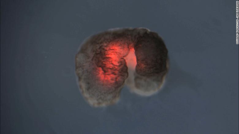
The organization and function of microtubules change dramatically during the cell cycle. At the onset of mitosis, a radial array of microtubules is broken down and reorganized into a bipolar spindle. This event requires changes in the dynamic behavior of individual microtubules. Through the use of Xenopus laevis egg extracts, a number of proteins affecting microtubule behavior have been identified. Recently, progress has also been made towards understanding how the activities of such microtubule-affecting proteins are regulated in a cell cycle-dependent manner. It is hoped that understanding how microtubule behavior is controlled during the cell cycle in vitro may illuminate the role of microtubule dynamics in various cellular processes.
Organelles are specialized structures that perform various jobs inside cells. The term literally means “little organs.” In the same way organs, such as the heart, liver, stomach, and kidneys, serve specific functions to keep an organism alive, organelles serve specific functions to keep a cell alive. May 23, 2019
https://www.researchgate.net/public...and_between_large_Xenopus_egg_asters/downloadThe cleavage furrow in Xenopus zygotes is positioned by two large microtubule asters that grow out from the poles of the 1st mitotic spindle. Where these asters meet at the midplane they assemble a disc-shaped interaction zone consisting of anti-parallel microtubule bundles coated with chromosome passenger complex (CPC) and centralspindlin which in.. Download full-text
During anaphase, a microtubule-containing structure called the midzone forms between the segregating chromosomes. The midzone is composed of an antiparallel array of microtubules and numerous microtubule-associated proteins that contribute to midzone formation and function. In many cells, the midzone is an important source of signals that specify the location of contractile ring assembly and constriction. The midzone also contributes to the events of anaphase by generating forces that impact chromosome segregation and spindle elongation; some midzone components contribute to both processes.
https://www.researchgate.net/public...indle_midzone_in_vertebrate_cells_at_a_glanceThe results of recent experiments have increased our understanding of the importance of the midzone, a microtubule array that has often been overlooked. This Journal of Cell Science at a Glance article will review, and illustrate on the accompanying poster, the organization, formation and dynamics of the midzone, and discuss open questions for future research.
That's a very interesting read, if you're into octopuses. (I've read it.) I don't regard it as a history of consciousness. Also, I don't see how this connects to the thread topic. I don't recall the book mentioning microtubules, for example.An essential history of the evolution of consciousness.
That's odd, I read a clear narrative of the evolution from fundamental chemical response mechanisms to ever greater neural complexity and sensory acuity that must have led to the emergence of conscious awareness of self and environment.That's a very interesting read, if you're into octopuses. (I've read it.) I don't regard it as a history of consciousness. Also, I don't see how this connects to the thread topic. I don't recall the book mentioning microtubules, for example.
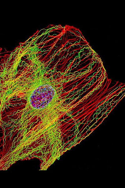
Microtubules may be the brains of the cell, particularly neurons—operating like a computerized Lego set. They are large complex scaffolding molecules that work closely with the two other rapidly changing structural molecules, actin and intermediate filaments, to provide structure for the entire cell including the spatial placement of organelles.
In neurons, microtubules respond instantly to mental events and constantly build and take down elaborate structures for the rapidly changing axons and dendrites of the synapses. Some think that microtubules are quantum computers and the seat of consciousness. Their lifestyle is quite remarkable.
A previous post described elaborate functions along the neuron’s axon including special tagging of cargoes that are transported by distinct motors with complex ancillary molecules for each type of transport.

Microtubules are the basic structural elements for cell division. The centromere is a key structure holding chromosomes together. It connects with the kinetochore where microtubule based spindle fibers attach to the chromosomes. Centrioles produce microtubules that orchestrate the rearrangement and sorting of the DNA during the extremely elaborate process of cell division.
Complex arrangements of microtubules direct and pull all the elements of the division process through multiple phases. The structure for this process is considered the most complex machine ever discovered in nature and is based on microtubule actions.
Are Microtubules the Brain of the Neuron?Microtubules are critical for the neuron’s migration; for the polar structure of the dendrite, soma and axon; and for stem cell’s determination of the type of cell a neuron will become. They are highways for long distance transport of materials and organelles. They are dynamic and changeable with elaborate mechanical functions. They control the signaling at local regions of the extremely long axon.
much, much more....... do take the time to read this comprehensive review of microtubule function.The actions of microtubules are fantastically complex. They respond instantly in a vast amount of different ways to mental events in thousands of places throughout the neuron at once. How is this directed? How can anyone think that this is a random process? How can anyone not see that mental events direct these fantastically complex processes?

Life has been generally considered to be a cell or group of cells, with metabolic functions and the ability to reproduce. It has not been at all clear how to define consciousness or intelligence.
But, now we find that much of a cell’s function is to converse with other cells or internally signal among its own components. This back and forth directed communication now has to be included in a new definition of life and perhaps might be related to intelligence throughout evolution.
https://jonlieffmd.com/category/blog/cellular-intelligence-blogEverywhere we look, there are ubiquitous conversations among all cells in all organisms. Life might come down to the ability to converse, which is another way of saying life is based on the ability to transfer meaningful biological information.
Can you find any mention of the word "microtubules" in the book? I don't recall any.IMO it showed the awesome versatility in evolutionary processes as long as microtubules (neurons) are present.
Eukaryotic lifeforms have lots of common denominators, so why you would single out microtubules would be a mystery, were it not for your record of fanboy devotion to them.I don't understand why you still reject the notion that microtubules are the single common denominator in all Eukaryotic lifeforms, both in the cellular cytoplasm and the neural networks of the body and brain, and even in plants for photosynthesis and distribution of energy throughout the leaves and stems.
Penrose's interest in them was put forward at least 30 years ago, now, as a tentative hypothesis in a book that was mostly about something else.Penrose was particularly interested in very simple single celled organism that did not have neural networks but did have cytoskeletons (microtubules) and were able communicate, coordinate, effect transport and navigation, mitosis and cell divisio, memory storage and recognition. If that is enough to pique a Nobel Prize winning scientist, who feels qualified to dismiss his curiosity as a waste of time?
The term neuron always includes microtubules. They are the heart of neurons and all cells for that matter. MT make up the cytoskeleton of ALL Eukaryotic organisms. Look it up!Write4U:
Can you find any mention of the word "microtubules" in the book? I don't recall any.
Name the functional common denominators in ALL Eukaryotic organisms that are present in the trillions of dynamic data processing networks throughout the entire body, effectively making the entire body an extension of the central processor brain.Eukaryotic lifeforms have lots of common denominators, so why you would single out microtubules would be a mystery, were it not for your record of fanboy devotion to them.
Penrose was astounded that single cells without any neural network could still communicate via the cytoskeleton (microtubules) itself. It is that remarkable ability in single celled organisms that raised the question if microtubules are suitable for quantum processing of qubits. This is how the collaboration with Hameroff came about.Penrose's interest in them was put forward at least 30 years ago, now, as a tentative hypothesis in a book that was mostly about something else.
And that is where you are wrong, due to willful refusal to put any serious research into the subject.I have not said that the study of microtubules is a waste of time, or anything like that. I have said that there's no good evidence for wild claims such as your ones that they are involved in memory, consciousness, recognition, coordination, etc. The vast majority of the sources you spam to this thread have nothing to do with any of that, either, even though this is your central thesis.

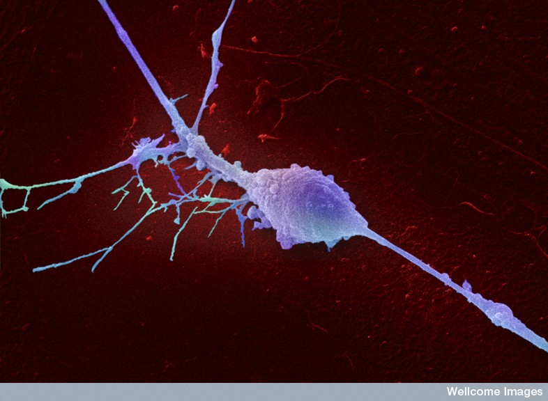
MTs are shown as light purple tubes, F-actin as red lines. MAPs are represented in blue, kinases in yellow and MAP-interacting proteins in purple. Guidance cue receptors are in brown. Guidance-evoked responses are represented in green (attraction), red (repulsion) and orange (pause) arrows. MT advance and retraction are represented with green and red arrowheads, respectively.
Cite , Download full-textDuring the establishment of neural circuitry axons often need to cover long distances to reach remote targets. The stereotyped navigation of these axons defines the connectivity between brain regions and cellular subtypes. This chemotrophic guidance process mostly relies on the spatio-temporal expression patterns of extracellular proteins and the s... more...
That's a "no", then. As I thought.The term neuron always includes microtubules.
As I understand it, they are structural components.They are the heart of neurons and all cells for that matter. MT make up the cytoskeleton of ALL Eukaryotic organisms. Look it up!
You speak as if there are "dynamic data processing networks" throughout the entire body. What evidence do you have for data processing at the microtubule level (as opposed to the the neuronal level)? Anything?Name the functional common denominators in ALL Eukaryotic organisms that are present in the trillions of dynamic data processing networks throughout the entire body, effectively making the entire body an extension of the central processor brain.
It's not really my field of expertise, and I'm not that interested. I'm sure that researchers in the field will let us know if they ever discover any actual "data processing" or whatever.If you have access to Research Gate, check them out for serious and formal presentations of current state of research into the nanoworld of microtubules.
How odd. You accuse me of posting anything that has the word microtubule in it. Now you accuse me of posting about neurons that have microtubules in them but does not explicitly mention microtubules.That's a "no", then. As I thought.
Yes, you have an extremely limited understanding of microtubules and their importance.As I understand it, they are structural components.
I anticipated that question for a long time now, demonstrating that you have no clue about the functions microtubules perform.You speak as if there are "dynamic data processing networks" throughout the entire body. What evidence do you have for data processing at the microtubule level (as opposed to the neuronal level)? Anything?
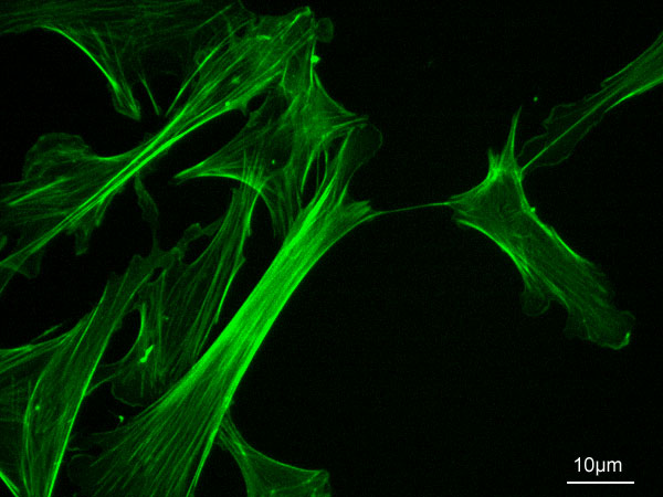
Microtubules (MTs) are long cylindrical structures of the cytoskeleton that control cell division, intracellular transport, and the shape of cells. MTs also form bundles, which are particularly prominent in neurons, where they help define axons and dendrites. 9 Aug 2018
Microtubules (MTs) are long cylindrical structures of the cytoskeleton that control cell division, intracellular transport, and the shape of cells.
MTs also form bundles, which are particularly prominent in neurons, where they help define axons and dendrites. MTs are bio-electrochemical transistors that form nonlinear electrical transmission lines.
However, the electrical properties of most MT structures remain largely unknown. Here we show that bundles of brain MTs spontaneously generate electrical oscillations and bursts of electrical activity similar to action potentials.
Under intracellular-like conditions, voltage-clamped MT bundles displayed electrical oscillations with a prominent fundamental frequency at 39 Hz that progressed through various periodic regimes. The electrical oscillations represented, in average, a 258% change in the ionic conductance of the MT structures. Interestingly, voltage-clamped membrane-permeabilized neurites of cultured mouse hippocampal neurons were also capable of both, generating electrical oscillations, and conducting the electrical signals along the length of the structure.
IntroductionOur findings indicate that electrical oscillations are an intrinsic property of brain MT bundles, which may have important implications in the control of various neuronal functions, including the gating and regulation of cytoskeleton-regulated excitable ion channels and electrical activity that may aid and extend to higher brain functions such as memory and consciousness.
MTs are unique components of the cellular cytoskeleton that form a wide variety of intracellular superstructures1. Highly polarized cells such as neurons, for example, present two structurally and
functionally distinct domains, namely a single long, thin axon and multiple shorter dendrites that either transmit or receive electrical signals, respectively. MT stability is at the center of the polarization process of neurons, which is fundamental to their development and plasticity, as well as the development of neurodegenerative diseases.
MTs form dense parallel arrays known as bundles in axons and dendrites, which are required for the growth and maintenance of neurites in neurons
Another important aspect of MT organization and function relies on their remarkable and not as well understood biophysical properties. MTs are highly charged electrically polarized polymers, where their αβ tubulin heterodimeric units have a high electric dipole moment
MTs are thought to generate oscillatory electric fields at expense of elasto-electrical vibrations
12, which may explain our findings that electrically-stimulated MTs behave as biological transistors behaving as sophisticated nonlinear transmission lines, capable of supporting the amplification and axial transfer of electrical signals 13,14,15,16,17,18. Within the cytoplasm MT-generated variable currents may contribute to the presence and modulation of large intracellular electric fields, which in turn, will help control cell function.
The above arguments support a potentially relevant role of electrical oscillations on brain MT bundles, which should be critical to neural function.
much more.......We recently reported that MT sheets sustain electrical oscillations19, which are driven by a permanent electrical polarization from local asymmetries in the ionic distributions between the intra- and extra-MT environments. Thus, the MT wall behaves as an electrical oscillator that produces oscillatory ionic currents with variable amplitude and periodicity depending on the driving force and ionic compositions, and is consistent with the periodic on-off switching of the nanopores. In this context, here we explored whether rat brain MT bundles do have electrical properties consistent with electrical oscillators. Our findings indicate that rat brain MT bundles generate a wide variety of endogenous electrical oscillations. Interestingly, the cytoskeleton of cultured adult mouse hippocampal neurons also supported electrical oscillations establishing an electrical role for brain MTs.
That is obvious.It's not really my field of expertise, and I'm not that interested.
Too bad you are not interested enough to read the state of knowledge about the role microtubules play in "data processing" at all levels and individual cells of all Eukaryotic organisms and in proto form in Prokaryotic organisms.I'm sure that researchers in the field will let us know if they ever discover any actual "data processing" or whatever.
I am sure you know that neurons are cells. Microtubules in the cytoplasm are what form and control the structure and behavior of cells.What evidence do you have for data processing at the microtubule level (as opposed to the neuronal level)? Anything?
Microtubule Structures Shape the Entire CellAxons can have as many as 100 bundles of microtubules in one axon cross section. There are many variations in these lattices with different types of stabilizing molecules, different orientations, and many different associated molecules and co factors. 29 Nov 2015
Microtubules and actin scaffolding direct all of this.When a neuron is being produced and while it migrates, it changes shape. Other shape changes occur frequently with new synapses both axons and dendrite spines (see post on Dendrite Spine Complexity). When a neuron migrates it produces a process in front, moves the nucleus to the front and then disassembles the process left in back.
The anchor in this process is the centrosome made of centrioles that are made of specific microtubules structures. It produces microtubule connections in the processes moving forward. The centrosome is the organizing center of the actions of microtubules. It is an organelle near the nucleus. Two centrioles at right angles are surrounded by a large protein mass. This very complex machine and directs cell division by pulling all elements of the division through many phases.
The centriole is a set of microtubules often arranged as a tubule with nine microtubules defining the circle. In some there are other microtubules in the center. (See post on the Primary Cilium for a discussion its centrioles). When centrioles connect, they do so at right angles and these pairs travel to opposite ends of the nucleus in the cell division process. Centrioles are involved in the organization of the mitotic spindle and in the completion of cytokinesis, or movement of the DNA in cell division.

But, centrosomes, made of centrioles, are, also, the critical way the neuron organizes the spreading and constantly changing microtubule structure. In fact, the centriole determines where the nucleus sits in the cell as well as organizing spatial structure of organelles in the cell (golgi, endoplasmic reticulum, etc). In cells with cilia and flagella, the central centriole determines where it will be. This mother centriole is, also, called the basal body as the starting point of the cell’s entire microtubule process.
Microtubules make a large structure that surrounds the entire nucleus in a cage. This cage extends from the centrosome around the nucleus and into the leading process. These microtubules orchestrate the neuron’s ability to migrate. The tubule structure then pulls the centrosome with the nucleus into the leading edge.
When the neuron becomes a specific type, microtubules take on very specific shapes and have to maintain these with unique stabilizing molecules. This involves very active transport of these stabilizing molecules by kinesin motors. How this is directed is not clear. It is possible that the centrosome and Golgi are involved. As the axon grows longer, many different stabilizing proteins are involved, such as +TIPs, interacting with actin, adhesion molecules, and the microtubules. At times an entire bundle of many microtubules are moved by mechanical forces from motors to allow for changes in shape. When injury to the axon occurs, microtubules are again critically involved in the rebuilding.
https://jonlieffmd.com/blog/are-microtubules-the-brain-of-the-neuronThe microtubules have many different roles in forming and stabilizing synapses. A previous post showed the dynamic changes of dendrite spines and the different shapes. This occurs through microtubule actions. The growth into the spines is regulated by factors like BDNF. These microtubules bring material for the changing spine shapes with special motors.