Write4U
Valued Senior Member
In furtherance of reference to:
Call-and-response circuit tells neurons when to grow synapses
Scientists explains signaling mechanism between astrocyte cells and neurons that regulates synaptic development, Date: October 25, 2021
Introduction
Rapidly accumulating physiological and genetic evidence establishes that the molecular diversity of synapses extends far beyond that envisioned by traditional classification schemes based solely on neurotransmitter identity. For instance, it is now clear that within each neurotransmitter category (e.g., glutamatergic, GABAergic, cholinergic) there is substantial diversity in the expression of many intrinsic synaptic proteins, including neurotransmitter transporters and receptors (Gupta et al., 2000; Häusser et al., 2000;taple et al., 2000;Cherubini and Conti, 2001; Craig and Boudin, 2001; Cull-Candy et al., 2001; Grabowski and Black, 2001;Huang and Bergles, 2004; Mody and Pearce, 2004; Grant, 2006 ). [/quote]
I find it remarkable that in this in-depth study there is no mention at all of microtubules which illustrates the fractured nature of scientific research. I have seen about five different descriptions of microtubules and their functions, but each study uses a different made-up name as if they were the first to discover the organelle.
When is a dedicated researcher going to combine all those separate studies and come up with a comprehensive and orderly compendium of the types and roles of microtubules (by any other name)?
I am too old and uninformed about neural science, but if I can find common denominators, then a knowledgeable student with access to libraries on formal neural research, should be able to put all this together and instead of asking the "hard question", be able to offer a comprehensive overview of "hard facts", that would surely lead to some "informed answers.

(A) A volume rendering of 60 serial sections (200 nm each) through the entire cortical depth, including portions of the striatum. While all subsequent experiments and analysis were performed on thinner, 70 nm sections, the thicker sections in this case have allowed us to collect a larger volume and to better visualize the extensive dendrites of pyramidal neurons. Synapsin (magenta), tubulin (blue), and YFP (green). Scale bar, 50 μm.
(B) A close up of layer 5 pyramidal neurons labeled with YFP.
(C–H) Zoomed-in view of layers 1 (C), 2/3 (D), 4 (E), 5a (F), 6a (G), and white matter and striatum (H).
Scale bar for (B)–(H), 10 μm.
See also Movie S1 for a more revealing rendering of the same image volume and Figure S1 for comparison of different synapsin antibodies.

Volume rendering from 20 sections, 70 nm each, from an array stained with 11 antibodies (Table S1, data set KDM-SYN-090416).
(A) Tubulin (blue), synapsin (magenta), YFP (green), and DAPI (gray) fluorescence.
(B–D) The boxed area in (A). DAPI (gray) and YFP (green). (B) Distribution of all presynaptic boutons as labeled with synapsin (magenta). (C) Distribution of VGluT1 (red), VGluT2 (yellow), and VGAT (cyan) presynaptic boutons. (D) Postsynaptic labels: GluR2 (blue), NMDAR1 (white), and gephyrin (orange) next to synapsin (magenta).
Scale bar 10 μm. See also Table S1 for sequence of antibody application.
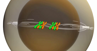
As described in post # 2211.Here we study the extent and the characteristics of self-organization using microtubules and molecular motors 2 as a model system. These components are known to participate in the formation of many cellular structures, such as the dynamic asters found in mitotic and meiotic spindles 3,4. Purified motors and microtubules have previously been observed to form asters in vitro 5.
Call-and-response circuit tells neurons when to grow synapses
Scientists explains signaling mechanism between astrocyte cells and neurons that regulates synaptic development, Date: October 25, 2021
Brain cells called astrocytes play a key role in helping neurons develop and function properly, but there's still a lot scientists don't understand about how astrocytes perform these important jobs. Now, a team of scientists led by Associate Professor Nicola Allen has found one way that neurons and astrocytes work together to form healthy connections called synapses. This insight into normal astrocyte function could help scientists better understand disorders linked to problems with neuronal development, including autism spectrum disorders. The study was published September 8, 2021, in the journal eLife.
"We know that astrocytes could play a role in neurodevelopmental disorders, so we wanted to ask: How are they playing a role in typical development?" says Allen, a member of the Molecular Neurobiology Laboratory. "In order to better understand the disorders, we first have to understand what happens normally."
https://www.sciencedaily.com/releases/2021/10/211025172108.htmSynapses form critical connections between neurons, allowing neurons to send signals and information throughout the body. Astrocyte cells play a role in synapse development by giving neurons directions, such as telling them when to start growing a synapse, when to stop, when to prune it back, and when to stabilize the connection.
Introduction
Rapidly accumulating physiological and genetic evidence establishes that the molecular diversity of synapses extends far beyond that envisioned by traditional classification schemes based solely on neurotransmitter identity. For instance, it is now clear that within each neurotransmitter category (e.g., glutamatergic, GABAergic, cholinergic) there is substantial diversity in the expression of many intrinsic synaptic proteins, including neurotransmitter transporters and receptors (Gupta et al., 2000; Häusser et al., 2000;taple et al., 2000;Cherubini and Conti, 2001; Craig and Boudin, 2001; Cull-Candy et al., 2001; Grabowski and Black, 2001;Huang and Bergles, 2004; Mody and Pearce, 2004; Grant, 2006 ). [/quote]
Until synapse molecular diversity is properly fathomed, it is likely to be a troublesome source of variability in physiological and neurodevelopmental experimentation.
https://www.cell.com/cms/10.1016/j....3799f533-26b8-40c5-ab6e-85bba89596da/mmc6.mp4Conversely, a systematic understanding of synapse diversity (i.e., the synaptome) is likely to provide valuable new perspectives on the organization of synaptic circuitry (i.e., the connectome), its development, plasticity and disorders. It is easy to envision, for instance, that a potential catalog of molecular synapse types (Grant, 2007) would help explorations of the synaptic basis of specific memory or disease processes to focus more fruitfully on specific synapse subpopulations.
I find it remarkable that in this in-depth study there is no mention at all of microtubules which illustrates the fractured nature of scientific research. I have seen about five different descriptions of microtubules and their functions, but each study uses a different made-up name as if they were the first to discover the organelle.
When is a dedicated researcher going to combine all those separate studies and come up with a comprehensive and orderly compendium of the types and roles of microtubules (by any other name)?
I am too old and uninformed about neural science, but if I can find common denominators, then a knowledgeable student with access to libraries on formal neural research, should be able to put all this together and instead of asking the "hard question", be able to offer a comprehensive overview of "hard facts", that would surely lead to some "informed answers.

(A) A volume rendering of 60 serial sections (200 nm each) through the entire cortical depth, including portions of the striatum. While all subsequent experiments and analysis were performed on thinner, 70 nm sections, the thicker sections in this case have allowed us to collect a larger volume and to better visualize the extensive dendrites of pyramidal neurons. Synapsin (magenta), tubulin (blue), and YFP (green). Scale bar, 50 μm.
(B) A close up of layer 5 pyramidal neurons labeled with YFP.
(C–H) Zoomed-in view of layers 1 (C), 2/3 (D), 4 (E), 5a (F), 6a (G), and white matter and striatum (H).
Scale bar for (B)–(H), 10 μm.
See also Movie S1 for a more revealing rendering of the same image volume and Figure S1 for comparison of different synapsin antibodies.

Volume rendering from 20 sections, 70 nm each, from an array stained with 11 antibodies (Table S1, data set KDM-SYN-090416).
(A) Tubulin (blue), synapsin (magenta), YFP (green), and DAPI (gray) fluorescence.
(B–D) The boxed area in (A). DAPI (gray) and YFP (green). (B) Distribution of all presynaptic boutons as labeled with synapsin (magenta). (C) Distribution of VGluT1 (red), VGluT2 (yellow), and VGAT (cyan) presynaptic boutons. (D) Postsynaptic labels: GluR2 (blue), NMDAR1 (white), and gephyrin (orange) next to synapsin (magenta).
Scale bar 10 μm. See also Table S1 for sequence of antibody application.
https://www.cell.com/neuron/fulltext/S0896-6273(10)00766-X?_returnIn addition to the YFP labeled neurons, the apical dendrites of pyramidal cells not expressing YFP are evident from tubulin immunostaining of their core microtubule bundles (Figure 1D).
Last edited:

/media/img/posts/2016/11/QBrain_615/original.png)


