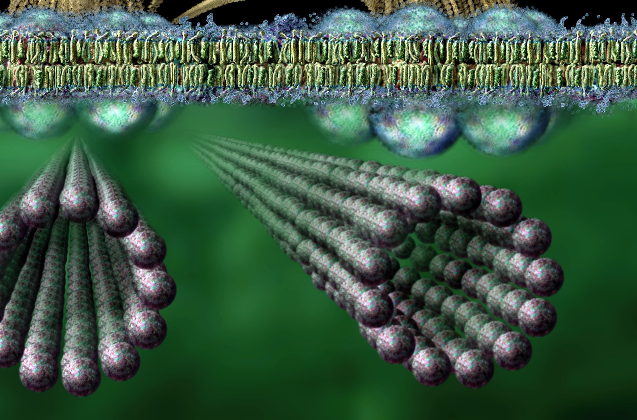
The "tau tangles" are not the ends of the microtubules. They are shown in blue after breaking off from the microtubule which is extending from the axon, showing the catastrophe in process, with the "tau particles" falling off the microtubule. The illustration identifies all the parts of the process, by NAME!!!!!!!
Put on your reading glasses.
That's nice.
But I was not addressing the image.
I was addressing the manner in which you altered a quote to make it appear as though it was talking about microtubules when it clearly originally was not.
You inserted "(microtubules)" in bold, altered that sentence, and the manner in which you did it gave the appearance that it was talking about microtubules. It was not.
The manner in which you post and reply is deliberately deceptive and dishonest.
Your first comment in that post alluded directly to microtubules:
Further confirmation of microtubule catastrophe in Alzheimer patients.
You then deliberately altered that quote by inserting "(microtubules)" after these words from the quote "that many of the fibers extending from the ends of the nerve cells "...
The quote I am addressing and the manner in which you deliberately misrepresented that quote by altering it to suit your narrative, is what is deceptive.
Not to mention the fact that you gave the impression that the article was about microtubules, or "microtubule catastrophe", when it clearly was not.. Just as your use of "microtubules catastrophe" in the context that you are using that term is deceptive.
Let me be clear, Write4U..
You altered a quote from an article and made it appear to be something else altogether..
It is blatantly dishonest and deceptive.
No, no, it clarifies any confusion in identification. I did that for your benefit, but as with the problems you had in separating microtubules from neurons, you're misinterpreting the use of the term "nerve cells". I merely made a clarification. You're not paying attention.
It is the microtubule that is disintegrating, not the nerve cell itself. The tau proteins fall off the microtubule, not the nerve cell, and form the tau tangles at the very bottom of the illustration. There is one "tangle" at the bottom left and another "tangle" at the bottom right part of the illustration.
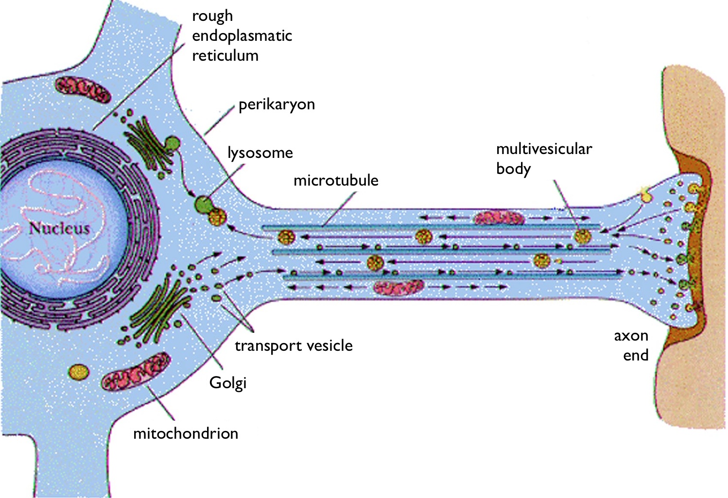
The picture at upper left is a healthy neuron and one of its microtubules with tau proteins assisting in growth of the microtubule.
Note: upper left says "healthy neuron", the lower right picture says "diseased neuron" at top and "disintegrating microtubule" at bottom.
It also shows the discoloration of the stressed neural cell itself, producing the plaque, perhaps?
Pay closer attention. You'll won't look so confused as you appear now.
Stop lying. I was addressing the quote. And it was quite clear that I was - given I posted the quote.. In its original format and the altered version that you posted.
Secondly, the image mentions microtubules because it is a part of the cell. You posted an altered quote, with a sentence about "microtubules catastrophe" and inserted "microtubules" in that quote to alter its meaning and context entirely.
This is basically what it comes down to.
I get it, you are all up about microtubules.
But when you resort to lying and being as dishonest as you have been, you simply come across as a zealot. And if you do it again, I'll issue you with an infraction and shut the thread down.
It's as simple as that.
If you cannot support your claims or argument without outright lying and that level of dishonesty, I'd suggest you refrain from making such claims.