Is consciousness to be found in quantum processes in microtubules?

Status
Not open for further replies.
Yet another example of the compound functional value provided by the microtubule and related filamental networks and functions.

Microtubule-Based Mitochondrial Dynamics as a Valuable Therapeutic Target in Cancer
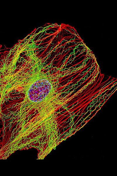
Simple Summary
Mitochondria are well known for being the powerhouses of the cell—whether the cell is normal or cancerous. Moreover, they can move, split, fuse themselves, or be eliminated via mitophagy with the help of the interplay between motor proteins and the cell scaffold—especially microtubules. The relationship between mitochondria, microtubules, and motor proteins is altered in cancer, and targeting this molecular machinery can offer a novel weapon in its treatment. In this paper, we review and summarize the state of the art of this approach.
Abstract
Mitochondria constitute an ever-reorganizing dynamic network that plays a key role in several fundamental cellular functions, including the regulation of metabolism, energy production, calcium homeostasis, production of reactive oxygen species, and programmed cell death. Each of these activities can be found to be impaired in cancer cells. It has been reported that mitochondrial dynamics are actively involved in both tumorigenesis and metabolic plasticity, allowing cancer cells to adapt to unfavorable environmental conditions and, thus, contributing to tumor progression.
The mitochondrial dynamics include fusion, fragmentation, intracellular trafficking responsible for redistributing the organelle within the cell, biogenesis, and mitophagy. Although the mitochondrial dynamics are driven by the cytoskeleton—particularly by the microtubules and the microtubule-associated motor proteins dynein and kinesin—the molecular mechanisms regulating these complex processes are not yet fully understood.
More recently, an exchange of mitochondria between stromal and cancer cells has also been described. The advantage of mitochondrial transfer in tumor cells results in benefits to cell survival, proliferation, and spreading. Therefore, understanding the molecular mechanisms that regulate mitochondrial trafficking can potentially be important for identifying new molecular targets in cancer therapy to interfere specifically with tumor dissemination processes.
1. Introduction
The cytoskeleton is a dynamic and interconnected network of filaments composed of structural and regulatory proteins that play a key role in all fundamental cellular processes, such as shape retention, motility, division, and intracellular transport of proteins and organelles [1,2]. Therefore, it is not surprising that alterations in cytoskeletal function can contribute to the onset and progression of cancer [3]. The three main types of filament that characterize the cytoskeleton are microfilaments, microtubules, and intermediate filaments [4]. Several ultrastructural analyses have shown that the cytoskeletal filaments interact directly or indirectly with the plasma membrane and various intracellular organelles [5].
Microtubules have a distinct polarity that is critical for their biological function. Tubulin polymerizes end-to-end; therefore, in an MT, one end will have the α-subunits (minus) exposed, while the other end will have the β-subunits (plus) exposed [7].
MTs are essential in many vital cellular processes, such as structural support, mitosis, chromosome segregation during meiosis, and intracellular transport of vesicles and organelles such as mitochondria [1,2,7]. In particular, to facilitate the movement of vesicles and mitochondria along their tracks, MTs recruit motor proteins via acetylation on lysine 40 of α-tubulin [8,9,10,11]. Microtubule-associated motor proteins include kinesin and dynein, which carry their cargo to the minus and plus ends of the microtubules, respectively [12,13]. In vitro studies have revealed that the loss of acetylated residues in MTs reduces the interaction of kinesin with tubulin, with a subsequent decrease in cell motility [10].
MTs have been an ideal target in antineoplastic therapy for many years, as they are the main components of the mitotic spindle. In addition, these filaments, together with motor proteins, play a fundamental role in the mitochondria’s structural and functional organization, including morphology, dynamics, motility, and distribution (Figure 1) [14].
upload_2022-8-13_3-43-49.png

Figure 1
Microtubule-dependent mitochondrial dynamics: Through the balance between fusion/fission and biogenesis/mitophagy, mitochondrial dynamics represent a central process in the bioenergetic adaptation and metabolic plasticity of cancer cells. The balance between biogenesis and mitophagy regulates the number of mitochondria and their quality. The fusion process helps to increase mitochondrial metabolism and to limit mitophagy and apoptosis, while the fission process allows the spatial redistribution of mitochondria in areas of the cell with greater energy and metabolic needs, favoring cell spreading and metastases.

https://www.ncbi.nlm.nih.gov/pmc/articles/PMC8616325/
 
Last edited:
This article on the self-correcting abilities of microtubules.

Abstract
The orientation of cell polarity depends on the position of the centrosome, the main microtubule-organizing center (MTOC). Microtubules (MTs) transmit pushing forces to the MTOC as they grow against the cell periphery. How the actin network regulates these forces remains unclear. Here, in a cell-free assay, we used purified proteins to reconstitute the interaction of a microtubule aster with actin networks of various architectures in cell-sized microwells. In the absence of actin filaments, MTOC positioning was highly sensitive to variations in microtubule length.
The presence of a bulk actin network limited microtubule displacement, and MTOCs were held in place. In contrast, the assembly of a branched actin network along the well edges centered the MTOCs by maintaining an isotropic balance of pushing forces. An anisotropic peripheral actin network caused the MTOC to decenter by focusing the pushing forces. Overall, our results show that actin networks can limit the sensitivity of MTOC positioning to microtubule length and enforce robust MTOC centering or decentering depending on the isotropy of its architecture.

Synopsis


embj2022111631-abs-0001-m.jpg


The structural and biochemical complexity of the cytoplasm has hampered analyses of the mechanical role of the actin network in positioning of the microtubule-organizing center (MTOC). Here, a reconstitution assay reveals that variations of actin network density in bulk or along the periphery can focus or balance pushing forces within the microtubules network.
more.....
https://www.embopress.org/doi/abs/10.15252/embj.2022111631
 
Which brings up the question if microtubules in external plant root systems have similar functions to microtubules in internal neural systems of surface organisms. Are plant roots a form of pseudopodia, walking vertically through the soil?
==========================

Microtubules in Purkinje cells may be the timekeepers in the brain.

Internal Clocks, mGluR7 and Microtubules: A Primer for the Molecular Encoding of Target Durations in Cerebellar Purkinje Cells and Striatal Medium Spiny Neurons
S. Aryana Yousefzadeh1, Germund Hesslow2, Gleb P. Shumyatsky3 and Warren H. Meck1*
  • 1Department of Psychology and Neuroscience, Duke University, Durham, NC, United States
  • 2Department of Experimental Medical Science, Lund University, Lund, Sweden
  • 3Department of Genetics, Rutgers University, Piscataway, NJ, United States
The majority of studies in the field of timing and time perception have generally focused on sub- and supra-second time scales, specific behavioral processes, and/or discrete neuronal circuits. In an attempt to find common elements of interval timing from a broader perspective, we review the literature and highlight the need for cell and molecular studies that can delineate the neural mechanisms underlying temporal processing.
Moreover, given the recent attention to the function of microtubule proteins and their potential contributions to learning and memory consolidation/re-consolidation, we propose that these proteins play key roles in coding temporal information in cerebellar Purkinje cells (PCs) and striatal medium spiny neurons (MSNs).
The presence of microtubules at relevant neuronal sites, as well as their adaptability, dynamic structure, and longevity, makes them a suitable candidate for neural plasticity at both intra- and inter-cellular levels. As a consequence, microtubules appear capable of maintaining a temporal code or engram and thereby regulate the firing patterns of PCs and MSNs known to be involved in interval timing. This proposed mechanism would control the storage of temporal information triggered by postsynaptic activation of mGluR7. This, in turn, leads to alterations in microtubule dynamics through a “read-write” memory process involving alterations in microtubule dynamics and their hexagonal lattice structures involved in the molecular basis of temporal memory.

more..... https://www.frontiersin.org/articles/10.3389/fnmol.2019.00321/full
 
To add some additional perspective.

What makes microtubules the only candidate for the job is the fact that they do the dynamic processing of data and create the “action potentials” that cause the dynamics in living organisms, which start with the cilia and flagella providing motility in single-celled organisms like the Paramecium and transport all sensory data to the brain for cognitive processing in brained organisms.

We keep talking about “small” and inconsequential because microtubules are ~ 20 nm.
Allow me to put this in proper perspective.

Transmitting fibers in the brain: Total length and distribution of lengths
The human brain’s approximately 86 billion neurons are probably connected by something like 850,000 km of axons and dendrites. Of this total, roughly 80% is short-range, local connections (averaging 680 microns in length), and approximately 20% is long-range, global connections in the form of myelinated fibers (likely averaging several centimeters in length).
And this is just in the brain alone! If you want to add the total length of the body’s neural network then we end up with something like ;

The nervous system: more than 90,000 miles of sensations!
The structure of the nervous system
The nervous system allows our bodies to perceive sensations, to think and to perform all of our movements, both voluntary and involuntary. It is composed of the brain, the spinal cord and the nerves.
Anatomically speaking, the nervous system is comprised of the central nervous system (the brain and the spinal cord, which are the interpretation and command centers), and the peripheral nervous system, which is composed of the nerves (the transmission network).
http://www.ikonet.com/en/visualdictionary/static/us/the_nervous_system

And the actual components inside neurons doing the actual work are…microtubules, also called the data transport highways much like the copper wiring and switches in electric cables and data processors inside a computer or in a home's electrical wiring system that create electrical fields created by the system's dynamics.


The Microtubule Cytoskeleton at the Synapse
Julie Parato1,2 and Francesca Bartolini1,*

Abstract

In neurons, microtubules (MTs) provide routes for transport throughout the cell and structural support for dendrites and axons. Both stable and dynamic MTs are necessary for normal neuronal functions. Research in the last two decades has demonstrated that MTs play additional roles in synaptic structure and function in both pre- and postsynaptic elements.
Here, we review current knowledge of the functions that MTs perform in excitatory and inhibitory synapses, as well as in the neuromuscular junction and other specialized synapses, and discuss the implications that this knowledge may have in neurological disease
more....https://www.ncbi.nlm.nih.gov/pmc/articles/PMC8089059/

There is no comparable common data processing system in all of Eukaryotic life. Why is this so hard to accept?
 
Last edited:
There is no comparable common data processing system in all of Eukaryotic life. Why is this so hard to accept?
Because nothing you post actually talks about data processing in microtubules.

Microtubules are structural elements of cells, from what I can gather. Your most recent quote describes them as providing "routes for transport throughout the cell and structural support..."

You might as well claim that highways for cars are "data processors". Highways perform similar functions to microtubules, as far as I can tell.
 
Because nothing you post actually talks about data processing in microtubules.
Then you have not bothered to read any of the links I posted that describe in detail the electrochemical information microtubules process and why it is important that they act as variable resistors or potentiometers.

electronic-coil-ferrite-core-and-blue-small-color-coded-resistors-on-red-copper-wire-cylindric-electromagnetic-inductor-pcb-detail-of-cable-tv-amp-2JA4YF7.jpg

Why do we use these structures in electronics?


This is what natural selection found sufficiently versatile in function to produce by the trillions throughout the body and connected by 1000 trillion dynamic synapses in the brain. I don't think the brain needs much structural support, but it is designed to process enormous numbers of electrochemical data. This seems obvious to me.
Tubulin_Infographic.jpg

Microtubules play an important role in a number of cellular processes. They are involved in maintaining the structure of the cell and, together with microfilaments and intermediate filaments, they form the cytoskeleton. They also make up the internal structure of cilia and flagella. They provide platforms for intracellular transport and are involved in a variety of cellular processes, including the movement of secretoryvesicles, organelles, and intracellular macromolecular assemblies.[5] They are also involved in cell division (by mitosis and meiosis) and are the main constituents of mitotic spindles, which are used to pull eukaryotic chromosomes apart.

Contents
much more...... https://en.wikipedia.org/wiki/Microtubule#

continued......
 
.......continued
Microtubules are structural elements of cells, from what I can gather. Your most recent quote describes them as providing "routes for transport throughout the cell and structural support..."
Seems to me you are hung up on the term "cytoskeleton", but how about "cytoplasm", how about "neurons", how about "synaptic connections"? How about intra-cellular data processing? How about inter-cellular data processing?

Cellular Intelligence
Are Microtubules the Brain of the Neuron

Microtubules may be the brains of the cell, particularly neurons—operating like a computerized Lego set. They are large complex scaffolding molecules that work closely with the two other rapidly changing structural molecules, actin and intermediate filaments, to provide structure for the entire cell including the spatial placement of organelles. In neurons, microtubules respond instantly to mental events and constantly build and take down elaborate structures for the rapidly changing axons and dendrites of the synapses.
Some think that microtubules are quantum computers and the seat of consciousness. Their lifestyle is quite remarkable. A previous post described elaborate functions along the neuron’s axon including special tagging of cargoes that are transported by distinct motors with complex ancillary molecules for each type of transport.


Microtubules are the basic structural elements for cell division. The centromere is a key structure holding chromosomes together. It connects with the kinetochore where microtubule based spindle fibers attach to the chromosomes. Centrioles produce microtubules that orchestrate the rearrangement and sorting of the DNA during the extremely elaborate process of cell division. Complex arrangements of microtubules direct and pull all the elements of the division process through multiple phases.
The structure for this process is considered the most complex machine ever discovered in nature and is based on microtubule actions.
A mere structural building block?
James R said:
You might as well claim that highways for cars are "data processors". Highways perform similar functions to microtubules, as far as I can tell.
Yes and what would happen if there were no highways for cars to transport "data" like food and medicine and building materials?
Microtubules are critical for the neuron’s migration; for the polar structure of the dendrite, soma and axon; and for stemcell’s determination of the type of cell a neuron will become. They are highways for long distance transport of materials and organelles. They are dynamic and changeable with elaborate mechanical functions. They control the signaling at local regions of the extremely long axon.
The control of the microtubule function is extremely complex, establishing and maintaining the architecture of the neuron. The control is distributed throughout the cell, which brings up the question of where the central control is to respond to mental events in such detailed and complex ways.
They are highly regulated in terms of the number, length, allocation, exact positioning and placement. Abnormalities in any of these functions leads to brain disease. Mutations in the proteins that hold the microtubules in place, such as tau are critical for the development of major neurodegenerative diseases (see post). The motors that move material along the microtubule highways—dyneins and kinesins—and regulators of these motors—dynactins—can produce other brain diseases. Defective transport in the axon is associated with ALS, Alzheimer’s, and multiple sclerosis.
https://jonlieffmd.com/blog/are-microtubules-the-brain-of-the-neuron

And if microtubules were not involved in brain function, why do defective microtubules produce brain disease?

OTOH, we can also ask why is it that healthy microtubules produce healthy brain function?
Are they instrumental in the emergence of consciousness itself? If not why not? Is there a better candidate?
 
Last edited:
Write4U:

Perusing your last couple of posts, the thing that stands out to me the most is the repeated use of the word "structure" to describe the role that microtubules play. They are structural elements.

There's hardly any mention at all of anything remotely resembling "data processing".

Why do you keep talking around the issue rather than trying to address it?
 
Write4U:

Perusing your last couple of posts, the thing that stands out to me the most is the repeated use of the word "structure" to describe the role that microtubules play. They are structural elements.
That is a false staement. The structural part is the least connected with data transfer. Why do you insist on bringing that up as the only function microtubules perform when I repeatedly cite quotations that the structural part is only one of the many functions MT perform?

Is mitosis a structural function? Are neurons structural components? Is the brain subject to structural stresses?

There's hardly any mention at all of anything remotely resembling "data processing".
That is not true! There is abundant information about the other functions microtubules perform. You just refuse to look at it.
Early on you accused me of cherry-picking microtubule functions. Now you are cherry-picking only the structural function about cytoskeleton as if that is represented by
shopping
instead of
0ef42bc6-3449-4bd8-8fcc-d1f25e908bfd

Why do you keep talking around the issue rather than trying to address it?
It is you who keeps talking around the issue. I am not required to give an exact count and name of all the chemicals and electric "action potentials" that MTs generate in the course of their data processing.

You just refuse to look at the data processing part and I don't know why you purposely keep avoiding MT functions pertinent to data processing and their role in brain function.

I included the structural issue because it is one of the many roles MT play in the maintenance of homeostatis and growth function and pseudopodia.

Maybe a simple search on "microtubules in the brain" here on "sciforum search " You'll get 10 reference pages with synopsis.

Offhand, you may want to check out posts #2476 -2480, and actually read the quoted passages. I think there won't be much talk about "structure" in those posts.
 
Last edited:
I have posted this before but it bears repeating.

Bundles of Brain Microtubules Generate Electrical Oscillations

Abstract
Microtubules (MTs) are long cylindrical structures of the cytoskeleton that control cell division, intracellular transport, and the shape of cells. MTs also form bundles, which are particularly prominent in neurons, where they help define axons and dendrites. MTs are bio-electrochemical transistors that form nonlinear electrical transmission lines. However, the electrical properties of most MT structures remain largely unknown.
Here we show that bundles of brain MTs spontaneously generate electrical oscillations and bursts of electrical activity similar to action potentials.
Under intracellular-like conditions, voltage-clamped MT bundles displayed electrical oscillations with a prominent fundamental frequency at 39 Hz that progressed through various periodic regimes. The electrical oscillations represented, in average, a 258% change in the ionic conductance of the MT structures. Interestingly, voltage-clamped membrane-permeabilized neurites of cultured mouse hippocampal neurons were also capable of both, generating electrical oscillations, and conducting the electrical signals along the length of the structure.
Our findings indicate that electrical oscillations are an intrinsic property of brain MT bundles, which may have important implications in the control of various neuronal functions, including the gating and regulation of cytoskeleton-regulated excitable ion channels and electrical activity that may aid and extend to higher brain functions such as memory and consciousness.
https://www.nature.com/articles/s41598-018-30453-2#

The problem is that invasive brain research on living subjects is extremely difficult. This is why we must rely on anesthesiologists like Hameroff to inform us of the latest "discoveries" of microtubule brain functions.
 
Let's see. Some stuff about structure:
Microtubules (MTs) are long cylindrical structures of the cytoskeleton that control cell division, intracellular transport, and the shape of cells. MTs also form bundles, which are particularly prominent in neurons, where they help define axons and dendrites. MTs are bio-electrochemical transistors that form nonlinear electrical transmission lines. However, the electrical properties of most MT structures remain largely unknown.
Followed by a repeat of something that Write4U brought up earlier in the thread, which does not mention data processing:
Under intracellular-like conditions, voltage-clamped MT bundles displayed electrical oscillations with a prominent fundamental frequency at 39 Hz that progressed through various periodic regimes. The electrical oscillations represented, in average, a 258% change in the ionic conductance of the MT structures. Interestingly, voltage-clamped membrane-permeabilized neurites of cultured mouse hippocampal neurons were also capable of both, generating electrical oscillations, and conducting the electrical signals along the length of the structure.
Electrical oscillations are not data processing. The conduction of electric signals along a structure is not data processing.

Then we have another repeated quote from earlier in the thread:
Our findings indicate that electrical oscillations are an intrinsic property of brain MT bundles, which may have important implications in the control of various neuronal functions, including the gating and regulation of cytoskeleton-regulated excitable ion channels and electrical activity that may aid and extend to higher brain functions such as memory and consciousness.
Again, electrical oscillations are not data processing. The rest is speculation: microtubules might conceivably do this and might conceivably have implications for that and might aid this or that. Might, might, might. Or might not.

"Higher brain functions" do involve data processing, but there's no evidence here for any role for microtubules in that processing, other than perhaps as providing highways for "electrical oscillations" and "structures" that scaffold cells.
 
I came across a 2021 paper on nih.gov that reviews "the known theories discussing the essence of consciousness" and makes "an attempt to integrate these explanations" in "an interdisciplinary, integrated theoretical model" that is presented "in a manner intuitively understandable." (Różyk-Myrta, et al., 2021) The authors include the ‘Orch OR’ microtubule theory in their model and explain its place in the holistic concept of self. They attempt to provide viable mechanisms for many phenomena of consciousness; including ego, self awareness, imagery and qualia perceptions.

The paper is well written, footnoted, sourced and linked, with an informative glossary included. I found the essay fascinating and consider it the best summation yet encountered of current theses addressing the hard problem of consciousness through biology and physics. It may assist readers in comprehending what is meant by microtubules performing data processing and potentially being the sine qua non of consciousness, albeit as only one of several interrelated components.

Abstract

"The known theories discussing the essence of consciousness have been recently updated. This prompts an attempt to integrate these explanations concerning several distinct components of the consciousness phenomenon such as the ego, and qualia perceptions. Therefore, it is useful to consider the latest publications on the ‘Orch OR’ and ‘cemi’ theories, which assume that quantum processing occurs in microtubules and that the brain’s endogenous electromagnetic field is important. The authors combine these explanations with their own theory describing the neural circuits realizing imagery. They try to present such an interdisciplinary, integrated theoretical model in a manner intuitively understandable to people with a typical medical education. In order to do this, they even refer to intuitively understandable metaphors. The authors maintain that an effective comprehension of consciousness is important for health care professionals because its disorders are frequent medical symptoms in emergencies, during general anesthesia and in the course of cognitive disorders in elderly people. The authors emphasize the current possibilities to verify these theses regarding the essence of consciousness thanks to the development of functional brain imaging methods—magnetoencephalography, transcranial magnetic stimulation—as well as clinical studies on the modification of perceptions and feelings by such techniques as mindfulness and the use of certain psychoactive substances, especially among people with self-awareness and identity disorders."

Różyk-Myrta A, Brodziak A, Muc-Wierzgoń M., "Neural Circuits, Microtubule Processing, Brain's Electromagnetic Field-Components of Self-Awareness", National Library of Medicine: National Center for Biotechnology Information: Brain Sciences, 2021 Jul 25;11(8):984. doi: 10.3390/brainsci11080984. PMID: 34439603; PMCID: PMC8393322, https://www.ncbi.nlm.nih.gov/pmc/articles/PMC8393322/

* My apologies if this source has previously been referenced in this thread, I performed a few searches using key passages from the article and did not turn up anything - although I semi-regularly follow this thread and do not recall seeing it, that doesn't necessarily mean it isn't here already...
 
This seems like two comprehensive illustrations of the mechanics of neural data processing.

I'll just post the illustrations, but in the link they are accompanied by a sizable article with detailed explanations of the various functions.
upload_2022-9-20_18-26-46.png
Figure 2
Axon structure and key mechanisms of axon growth. The image shows a neuron which extends an axon into a zone of an attractive factor. Parallel bundles of MTs fill the axon and splay in the growth cone (plus end-out polarity indicated for some MTs by encircled + and - signs). F-actin networks are prominent in growth cones, and scarce but highly organized into evenly spaced rings in axons. Close-ups illustrate the following molecular mechanisms contributing to axon growth: (1) MT plus end-associated factors (for example, EB1, CLASP, type 13 kinesins, XMAP215; blue ellipses) regulate elongation and shrinkage of MTs (blue arrows); (3) molecular motors (orange Y structures) mediate cargo transport; (2,4) the MT severing protein katanin (scissors) generates MT fragments which are moved anterogradely through MT-sliding; (5) proteins that bind along MTs (for example, tau, MAP1B; brown L's) protect MTs from depolymerization or severing factors and organize MTs by cross-linking them and regulating their spacing; (6–8) F-actin networks influence MT behaviors through antagonizing MT advance into lamellipodia and filopodia via retrograde flow (red arrows), through forming structures that can be supportive (for example, radial bundles) or inhibitory (for example, transverse arcs), through MT cross-linkage (yellow stars), through contractile activity (not shown), or through F-actin clearance from protrusions (shown in 8). For detailed information see [3,23].

and
upload_2022-9-20_18-34-29.png
Figure 3
Structural aspects of tubulins and microtubules (MTs). (A) MTs grow through head-to-tail polymerization of α-/ß-tubulin heterodimers into protofilaments that arrange into hollow tubes through lateral interactions. The α-and ß-tubulin monomers need to be folded properly assisted by chaperones, they heterodimerize through longitudinal interactions (peach arrow), they bind GTP (of which GTP on ß-tubulin tends to undergo hydrolysis to GDP) and undergo a number of posttranslational modifications, including de-tyrosination (often followed by de-glutamylation resulting in ∆2-tubulin), acetylation, poly-amination, (poly-)glycylation and (poly-)glutamylation [11]. MTBPs primarily interact with the C-terminus of tubulins which sticks out from the MT surface. (B) The secondary structure of α- and ß-tubulin (color-coded as in A) showing the positions of ß-sheets (B1-10) and α-helices (H1-12; image modified from [24]); the borders and principal functions of the N-terminal, intermediate and C-terminal domains of ß-tubulin are indicated at the bottom and arrows indicate examples of posttranslational modification sites [11,21]. Positions of known dominant-negative mutations are shown below the two secondary structures [25]. Those mutations tested by Niwa and colleagues are highlighted in light blue, asterisks indicate those mutations that impair tubulin incorporation into MTs and blue arrowheads indicate the two charge-changing mutations in H12 [26].

https://www.ncbi.nlm.nih.gov/pmc/articles/PMC3846809/
 
Followed by a repeat of something that Write4U brought up earlier in the thread, which does not mention data processing:
What other dataprocessor in the body has dipolar (electrical) properties? In all the literature I have read, only the microtubule has been specifically identified as a dynamical dipolar coil, with ability to process electrical data and "action potentials" in a manner of a variable potentiometer.

Thoughts in the brain produce an electrical field that can be measured via EEG mechanics.

Electroencephalography
Diagnostic test


Description
Electroencephalography is a method to record an electrogram of the electrical activity on the scalp that has been shown to represent the macroscopic activity of the surface layer of the brain underneath. It is typically non-invasive, with the electrodes placed along the scalp. Wikipedia

Note; EEG use dipolar techniques for processing brain generated electrical data.

Dipole models for the EEG and MEG


Abstract

The current dipole is a widely used source model in forward and inverse electroencephalography and magnetoencephalography applications. Analytic solutions to the governing field equations have been developed for several approximations of the human head using ideal dipoles as the source model. Numeric approaches such as the finite-element and finite-difference methods have become popular because they allow the use of anatomically realistic head models and the increased computational power that they require has become readily available.
Although numeric methods can represent more realistic domains, the sources in such models are an approximation of the ideal dipole. In this paper, we examine several methods for representing dipole sources in finite-element models and compare the resulting surface potentials and external magnetic field with those obtained from analytic solutions using ideal dipoles.
https://pubmed.ncbi.nlm.nih.gov/12002172/

All informational data transport to the brain and emergent consciousness are provided by microtubules within the neural network.
 
Last edited:
Write4U:

I think you have a different idea of what "data processing" means than I do.

Suppose I connect a simple light bulb to a battery and it lights up because electrical current flows through the bulb. Would you say that the light bulb has the "ability to process electrical data"? I wouldn't.

The bulb takes an input (electrical energy) and produces an output (light), but there's no "processing" going on in the sense of manipulating old information to produce new information.

Similarly, a nerve carries an electrical impulse from one part of the body to another, but I wouldn't call that "data processing". A single nerve does not take in information and produce new information from it.

The brain as whole does do "data processing", in that it is able to take in information and manipulate it in various ways to produce novel information as an "output".

Do microtubules process data, or not?
 
My two cents, from what I can glean from those who seem to, individually or collectively, have a grip on advanced biology, neuroscience, electromagnetism, quantum physics, anesthesiology, psychiatry and a smattering of philosophy, is that the so called "data processing" is referring to decoherence, or collapse of, the alleged quantum entanglement in the microtubules, which then aids in triggering the firing of neurons, and so is viewed as analogous to "making a decision." (At least by some of these wizards)

Whether that meets the formal definition of "data processing" - well, my relevant microtubules have not yet collapsed on that issue :)
 
Suppose I connect a simple light bulb to a battery and it lights up because electrical current flows through the bulb. Would you say that the light bulb has the "ability to process electrical data"? I wouldn't.
In a quasi-intelligent mathematical universe I would call any conversion from pure energy into a coherent pattern "data processing".

Difference-between-data-and-information.png

https://byjus.com/biology/difference-between-data-and-information/

Perhaps I am too much of a romantic.

But IMO, microtubules are definitely data processors. They convert electrochemical data into "action potentials".
Moreover, the microtubule arrangement in "pyramidal neurons" act as data storage modules for "memory".

Of microtubules and memory: implications for microtubule dynamics in dendrites and spines
Erik W. Dent*
William Bement, Monitoring Editor

ABSTRACT
Microtubules (MTs) are cytoskeletal polymers composed of repeating subunits of tubulin that are ubiquitously expressed in eukaryotic cells. They undergo a stochastic process of polymerization and depolymerization from their plus ends termed dynamic instability. MT dynamics is an ongoing process in all cell types and has been the target for the development of several useful anticancer drugs, which compromise rapidly dividing cells.
Recent studies also suggest that MT dynamics may be particularly important in neurons, which develop a highly polarized morphology, consisting of a single axon and multiple dendrites that persist throughout adulthood. MTs are especially dynamic in dendrites and have recently been shown to polymerize directly into dendritic spines, the postsynaptic compartment of excitatory neurons in the CNS. These transient polymerization events into dendritic spines have been demonstrated to play important roles in synaptic plasticity in cultured neurons.
Recent studies also suggest that MT dynamics in the adult brain function in the essential process of learning and memory and may be compromised in degenerative diseases, such as Alzheimer’s disease. This raises the possibility of targeting MT dynamics in the design of new therapeutic agents.
more..... https://www.ncbi.nlm.nih.gov/pmc/articles/PMC5221613/

See also Purkinje neurons.
nerve8.jpg

Slide 7. Pyramidal and Purkinje cells
On the left, a photomicrograph of layer 3 of cerebral cortex showing two types of neurons: pyramidal and stellate. Pyramidal neurons (P) have a prominent apical dendrite (D). The axons (A) of pyramidal neurons exit the cortex and travel through white matter to the spinal cord, to the opposite cortex and to various nuclei in the brainstem. In contrast, stellate neurons (S) are local circuit neurons with synaptic connections to neighboring neurons.
On the right, a photomicrograph of a Purkinje cell in the cerebellum that was stained with the Golgi method, illustrating the cell body or soma (S) of the Purkinje cell and a portion of its extensive dendritic tree (D1, primary dendrite; D2, secondary dendrites).
https://web.indstate.edu/thcme/mmmoga/histology/slide114.html
 
Last edited:
Will wonders never cease? Microtubules are critically involved in the transport of transposons and transposons are a very interesting

Hijacking oogenesis enables massive propagation of LINE and retroviral transposons

SUMMARY
Although animals have evolved multiple mechanisms to suppress transposons, “leaky” mobilizations that cause mutations and diseases still occur. This suggests that transposons employ specific tactics to accomplish robust propagation. By directly tracking mobilization, we show that, during a short and specific time window of oogenesis, retrotransposons achieve massive amplification via a cell-type-specific targeting strategy.
Retrotransposons rarely mobilize in undifferentiated germline stem cells. However, as oogenesis proceeds, they utilize supporting nurse cells, which are highly polyploid and eventually undergo apoptosis, as factories to massively manufacture invading-products.
Moreover, retrotransposons rarely integrate into nurse cells themselves but, instead, via microtubule-mediated transport, preferentially target the DNA of the interconnected oocytes. Blocking microtubule-dependent intercellular transport from nurse cells significantly alleviates damage to the oocyte genome. Our data reveal that parasitic genomic elements can efficiently hijack a host developmental process to propagate robustly, thereby driving evolutionary change and causing disease.
In this study, we developed approaches to track transposon mobilization with spatiotemporal resolution during Drosophila oogenesis, which is a well-characterized process and has served as a critical model system to study the function of piRNA pathway (Mahajan-Miklos and Cooley, 1994; Siomi et al., 2011; Spradling, 1993).
As oogenesis proceeds, one germline stem cell gives rise to 15 supporting nurse cells and one oocyte. Although undergoing programmed cell death at the end of oogenesis, during the process of oocyte development, nurse cells produce the vast majority of cytoplasmic constituents/nutrients for oocyte from their highly polyploid genome (Mahajan-Miklos and Cooley, 1994; Spradling, 1993).
Here, we show that retrotransposons barely mobilize in germline stem cells. Upon differentiation, they utilize differentiated nurse cells to massively manufacture their invading products, but, seldom transpose into nurse cell DNA. Instead, via microtubule-mediated transport, retrotransposons selectively target the DNA of oocyte, the only ovarian cell that founds the next generation. Our data therefore demonstrate that retrotransposons exploit a hijacking tactic to robustly propagate in the host genome during oogenesis.
more......https://www.ncbi.nlm.nih.gov/pmc/articles/PMC6628338/
 
And now we are beginning to abstract several common properties in totally divergent species, into a theory about consciousness. It may even answer the age-old question of color perception.

The conscious intelligence of Cephalopoda shines a bright light on the ability of microtubule function in relation to conscious awareness, observation, and functional motor responses to environmental conditions, and by virtue of common functional denominators, extending to human intelligent responses to our environment.

Analysis of Microtubules in Isolated Axoplasm from the Squid Giant Axon
One obstacle to characterizing specific populations of neuronal microtubules is the complexity of nervous tissue. Separating neuronal microtubules from glial microtubules, dendritic microtubules from axonal or cell body microtubules is effectively impossible when using brain tissue as a source, so any studies on the biochemistry and biophysics of neuronal microtubules from brain reflect the properties of a mixed pool. The problem is compounded by the fact that a large fraction of neuronal tubulin is lost during standard preparations of brain tubulin and this population of stable microtubules has received little attention, despite representing more than 50% of axonal tubulin in mature neurons.
more...... https://www.ncbi.nlm.nih.gov/pmc/articles/PMC4460999/
______________________________________________________

Moreover, cuttlefish can copy colors, even as they only "see" black and white. This must be a function of the skin cells able to "recognize" the frequencies of colors. Think about that for a moment.

Cuttlefish: Wearing thoughts on the skin

original-1539849342.jpg


Cephalopods control camouflage by the direct action of their brain onto specialized skin cells called chromatophores, that act as biological color “pixels” on a soft skin display. Cuttlefish possess up to millions of chromatophores, each of which can be expanded and contracted to produce local changes in skin contrast. By controlling these chromatophores, cuttlefish can transform their appearance in a fraction of a second. They use camouflage to hunt, to avoid predators, but also to communicate.
A new technique is allowing researchers to study the inner workings of a cuttlefish brain by tracking colour changing cells in their skin. These cells are directly controlled by neurons extending from the brain. By monitoring the cells with high resolution cameras, researchers can track the activity tens of thousands of neurons at once for the first time.
Cuttlefish chromatophores are specialized cells containing an elastic sack of colored pigment granules. Each chromatophore is attached to minute radial muscles, themselves controlled by small numbers of motor neurons in the brain. When these motor neurons are activated, they cause the muscles to contract, expanding the chromatophore and displaying the pigment. When neural activity ceases, the muscles relax, the elastic pigment sack shrinks back, and the reflective underlying skin is revealed. Because single chromatophores receive input from small numbers of motor neurons, the expansion state of a chromatophore could provide an indirect measurement of motor neuron activity.
“This study opens up a large range of new questions and opportunities”, says Laurent. “Some of these concern texture perception and are relevant to the growing field of cognitive computational neuroscience; others help define the precise link between brain activity and behavior, a field called neuroethology; others yet help identify the cellular rules of development involved in tissue morphogenesis. Finally, this work opens a window into the brain of animals whose lineage split from ours over 540 million years ago. Cephalopod brains offer a unique opportunity to study the evolution of another form of intelligence, based on a history entirely independent of the vertebrate lineage for over half a billion years”.
https://www.mpg.de/12363924/1017-hirn-080434-elucidating-cuttlefish-camouflage

It is obvious that microtubule network and their related filaments in the brain and axoplasm of cephalopods play a major role in cognitive functions as they do in the microtubule network in the brain and cytoplasm of humans and indeed of all Eukaryotic life at varying levels of complexity.
 
Status
Not open for further replies.
Back
Top